Cartilage tissue engineering scaffold and preparation method thereof

A cartilage tissue and engineering technology, applied in the field of cartilage tissue engineering scaffolds and their preparation, can solve the problems of poor mechanical strength, poor mechanical strength of cartilage tissue engineering scaffolds, limited ability to recruit bone marrow mesenchymal stem cells, etc., to improve durability, The effect of strong cartilage tissue repair ability and convenient large-scale promotion and application

Active Publication Date: 2016-01-27
PEKING UNIV THIRD HOSPITAL
View PDF3 Cites 11 Cited by
  • Summary
  • Abstract
  • Description
  • Claims
  • Application Information

AI Technical Summary

Problems solved by technology

[0005] The prior art uses the above-mentioned materials such as proteins and carbohydrates to prepare cartilage tissue engineering scaffolds. Due to the poor mechanical strength of materials such as proteins and carbohydrates, the mechanical strength of the prepared cartilage tissue engineering scaffolds is relatively poor, so that the existing Technological cartilage tissue engineering scaffolds are less durable in clinical applications
At the same time, the cartilage tissue engineering scaffolds provided by the prior art have limited ability to recruit bone marrow mesenchymal stem cells, resulting in poor chondrogenic effect of the cartilage engineering scaffolds

Method used


Image

  • Cartilage tissue engineering scaffold and preparation method thereof
  • Cartilage tissue engineering scaffold and preparation method thereof
  • Cartilage tissue engineering scaffold and preparation method thereof

Examples


preparation example Construction

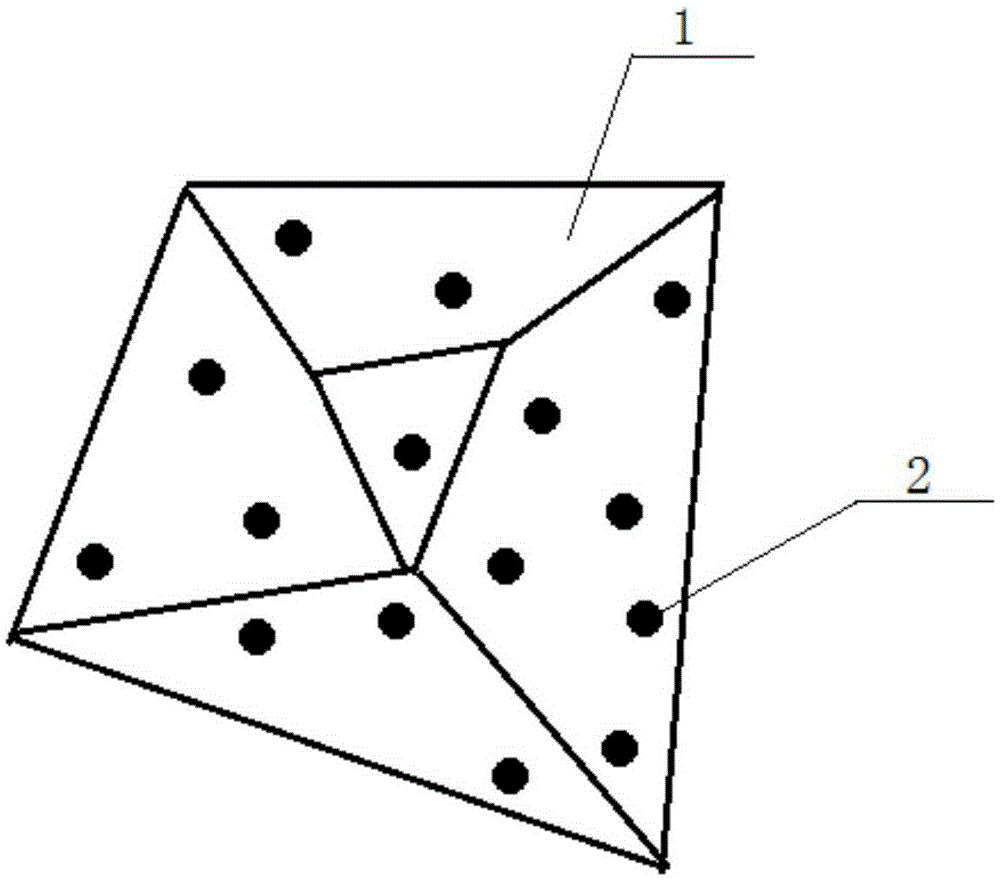
[0046] On the other hand, the embodiment of the present invention also provides the preparation method of the above-mentioned cartilage tissue engineering scaffold, comprising: preparing at least one decalcified cortical bone matrix microsphere 2 with a diameter of 500-800 μm; adding bone marrow mesenchymal stem cell affinity peptide modifying at least one surface of decalcified cortical bone matrix microsphere 2, and adding the decalcified cortical bone matrix microsphere 2 modified with bone marrow mesenchymal stem cell affinity peptide into the chitosan hydrogel solution, at 32-38°C The gelation reaction is carried out for 28-32 minutes, preferably 30 minutes, to obtain the cartilage tissue engineering scaffold.

[0047] Specifically, the preparation method of decalcified cortical bone matrix microspheres 2 is as follows: remove the soft tissue attached to the femur, such as muscle and fascia, obtain the cortical bone of the femur, and degrease and decalcify the cortical bon...

Embodiment 1

[0052] This embodiment provides a cartilage tissue engineering scaffold, comprising a chitosan hydrogel exoskeleton 1, and at least one decalcified cortical bone matrix microstructure coated with an E7 polypeptide on the surface of the chitosan hydrogel exoskeleton 1. ball 2.

[0053] The preparation method of the cartilage tissue engineering support provided by the present embodiment is:

[0054] Step 1. Preparation of decalcified cortical bone matrix microspheres

[0055] A urethane solution with a mass concentration of 20% was prepared, and a 4Kg New Zealand white rabbit was anesthetized with an excessive amount of urethane solution. The long bones of the limbs of the white rabbit were taken out, the muscles and fascia were removed to obtain the cortical bone of the femur, and then the cortical bone was placed in a mixed solution of methanol and chloroform and defatted for 48 hours. Wherein, the volume ratio of methanol and chloroform is 1:1. The degreased cortical bone ...

Embodiment 2

[0064] The purpose of this example is to illustrate the durability and mechanical properties of the cartilage tissue engineering scaffold provided in Example 1, and the in vitro degradation rate and equilibrium swelling ratio of the cartilage tissue engineering scaffold provided in Example 1 were determined. For the convenience of description, the cartilage tissue engineering scaffold provided in Example 1 is defined as the first scaffold 5 below. Wherein, the corresponding performances of the first comparison bracket 3 made of a single chitosan hydrogel and the second comparison bracket 4 composed of a single decalcified cortical bone matrix particle coated in a chitosan hydrogel are carried out the same The test process is used as a comparison.

[0065] The specific measurement process is as follows: firstly, the first comparison bracket 3 , the second comparison bracket 4 , and the first bracket 5 were respectively put into containers with a diameter of 2.5 mm and a height ...


PUM

PropertyMeasurementUnit
diameteraaaaaaaaaa
elastic modulusaaaaaaaaaa
pore sizeaaaaaaaaaa

Abstract

The invention discloses a cartilage tissue engineering scaffold and a preparation method thereof and belongs to the field of cartilage repair. The cartilage tissue engineering scaffold comprises a chitosan hydrogel outer skeleton and at least one decalcified cortical bone matrix microsphere coated in the chitosan hydrogel outer skeleton and with bone marrow mesenchymal stem cell affinity peptide modified on the surface, wherein diameter of each decalcified cortical bone matrix microsphere is 500-800 micrometers. The cartilage tissue engineering scaffold has high rheological property, easiness in plasticizing, biomechanical strength and performance of specifically gathering bone marrow mesenchymal stem cells, thereby being suitable for cartilage defect areas different in shape, lasting in effect and capable of acquiring more differentiated cartilage cells, has high cartilage tissue repair ability and is convenient for large-scale popularization and application.

Description

technical field [0001] The invention relates to the field of cartilage repair, in particular to a cartilage tissue engineering scaffold and a preparation method thereof. Background technique [0002] Cartilage tissue engineering is a technology that uses tissue engineering methods to construct cartilage composite tissues. The three basic elements in cartilage tissue engineering are: seed cells, degradable cartilage tissue engineering scaffolds, and cell growth regulators. Among them, the cartilage tissue engineering scaffolds play an important role in the process of cartilage repair. Cartilage tissue engineering scaffolds are mainly used to provide a three-dimensional space structure for the construction of chondrocytes, which is conducive to cell adhesion and proliferation, and provides a good growth environment for cell growth. [0003] At present, commonly used cartilage tissue engineering scaffolds are mainly prepared from natural biological scaffold materials, such as ...

Claims


Application Information

Patent Timeline
Patent Type & Authority Applications(China)
IPC IPC(8): A61L27/50A61L27/20A61L27/22A61L27/36
Inventor 敖英芳孟庆阳黄洪杰胡晓青代岭辉邵振兴
Owner PEKING UNIV THIRD HOSPITAL
Why Eureka
Try Eureka