Thermo-sensitive hydrogel and preparation method thereof

A temperature-sensitive hydrogel and collagen technology, applied in the field of temperature-sensitive hydrogel and its preparation, can solve the problems of short half-life of cytokines and the like

Inactive Publication Date: 2017-03-15
广东颜值科技有限公司
View PDF8 Cites 9 Cited by
  • Summary
  • Abstract
  • Description
  • Claims
  • Application Information

AI Technical Summary

Problems solved by technology

At present, it has been found that EpSCs can secrete dozens of growth factors closely related to the wound repair process, among which the more important ones are vascular endothelial growth factor (VEGF), epidermal growth factor (EGF), insulin-like growth factor-1 (IGF-1) , transforming growth factor-β (TGF-β), fibroblast gr...

Method used


Image

  • Thermo-sensitive hydrogel and preparation method thereof
  • Thermo-sensitive hydrogel and preparation method thereof
  • Thermo-sensitive hydrogel and preparation method thereof

Examples


Embodiment 1

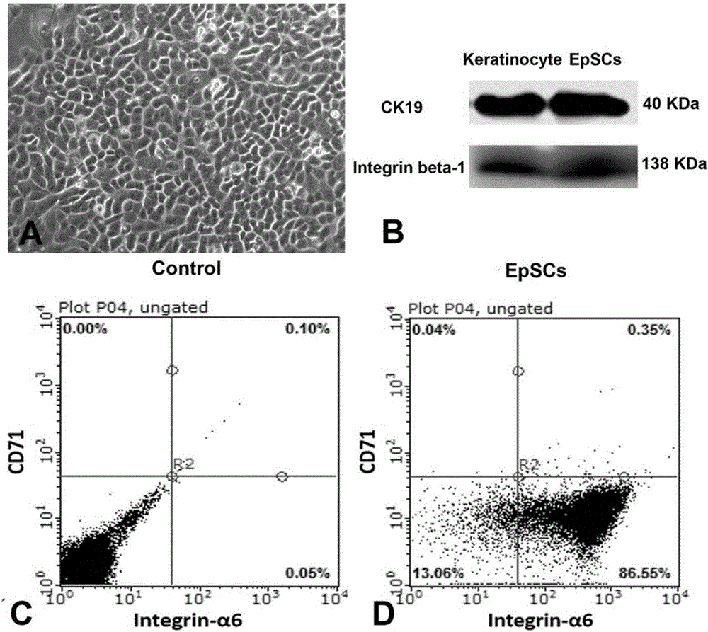
[0046] Example 1 Isolation and cultivation of epidermal stem cells EpSCs

[0047] experimental method:

[0048] 1) Rinse the freshly removed normal human skin with normal saline containing 100U / mL penicillin and streptomycin 10 times, cut into a skin piece of about 5mm×5mm, remove the subcutaneous fat tissue and separate the epidermis and dermis After obtaining the epidermal tissue, cut it into 1mm×1mm pieces again;

[0049] 2) Add 0.25% trypsin-PBS solution to the epidermis fragments to digest for 30 minutes, add EpSCs primary culture medium to stop digestion, and filter with a 100 μm filter after blowing; the filtrate is centrifuged at 1000r / min for 10 minutes, and the cell pellet is added to EpSCs primary culture medium (K-SFM medium, containing 10ng / ml of EGF, 2ng / ml of FGF and IGF-1, 30μg / ml of BPE, 0.05mM of CaCl 2 , 0.5μg / mL hydrocortisone, 100U / ml penicillin-streptomycin) to make 3×10 5 / ml of single-cell suspension, inoculated in a culture dish pre-coated with type...

Embodiment 2

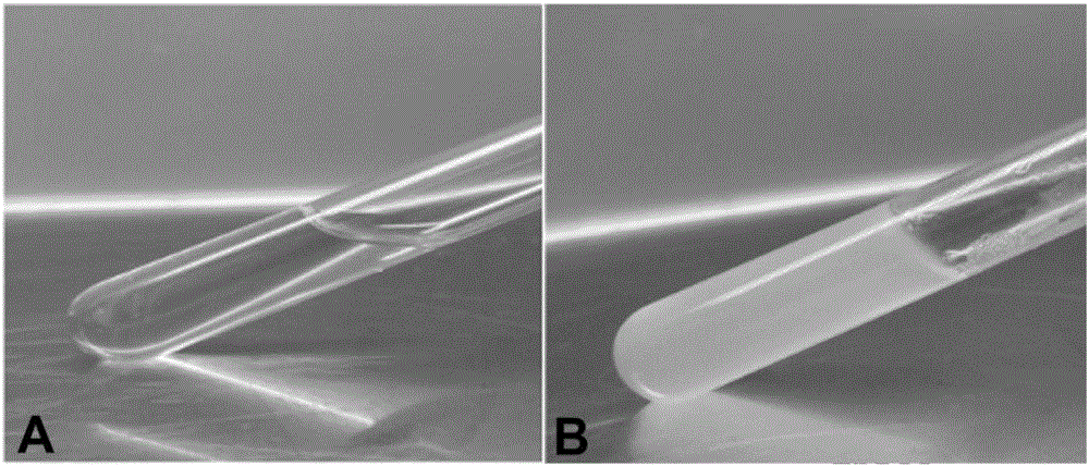
[0052] The preparation of embodiment 2 thermosensitive hydrogel

[0053] (1), preparation of epidermal stem cell conditioned medium: take the well-grown EpSC adherent cells of the 4th generation of culture, and when the fusion rate reaches 70-80%, the full amount of liquid is replaced with fresh EpSCs subculture medium. After 36 hours, collect the culture medium, centrifuge at 3000r / min for 30min to remove cell debris, filter and sterilize with a 0.22μm filter membrane, and use it as the EpSC conditioned medium;

[0054] (2), preparation of cytokine activity stabilizer: chitosan powder is dissolved in the hydrochloric acid solution of 0.1mol / L, makes the chitosan solution of 2% (W / V), then adds mannitol powder to make it The final concentration is 0.25%, then add polyethylene glycol powder to make the final concentration 1%, then add macromolecular hyaluronic acid with a molecular weight of 1.2-1.4 million to make the final concentration 0.5% (W / V), then add seaweed NaOH so t...

Embodiment 3

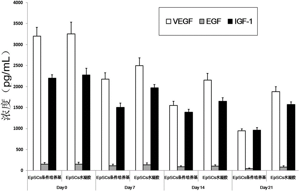
[0058] Example 3 Thermosensitive hydrogel maintains the activity of cytokines

[0059] (1) Put the temperature-sensitive hydrogel obtained in Example 2 in an environment of 4°C, take a portion on the 0th, 7th, 14th, and 21st day, squeeze out the leachate, and use the ELISA method to detect VEGF, EGF, and IGF-1 content;

[0060] (2) Dilute the EpSC conditioned medium obtained in step (1) of Example 2 to one time, place it in a 4°C environment, take a portion on the 0th, 7th, 14th, and 21st day, and use ELISA method to detect VEGF , EGF, IGF-1 content;

[0061] Test results such as image 3 As shown, it can be seen that compared with the conditioned medium, the thermosensitive hydrogel of the present invention can better maintain the concentration of cytokines VEGF, EGF, and IGF-1 within 21 days.


PUM


Abstract

The invention discloses a thermo-sensitive hydrogel. The thermo-sensitive hydrogel comprises the following raw materials: a cytokine activity stabilizer, an epidermal stem cell conditioned culture medium and a collagen cross-linking solution. The cytokine activity stabilizer is added into the epidermal stem cell conditioned culture medium, so that multiple cytokines secreted by EpSCs are stabilized, the half-life period of each of the multiple cytokines is prolonged, the effect of each of the multiple cytokines on trauma repair is prolonged, and further, wound healing is promoted. In addition, the invention discloses a preparation method of the thermo-sensitive hydrogel, which has the characteristics of being simple to operate, random in molding and capable of in-situ gelling; the prepared hydrogel is slightly-viscous and clear liquid under the condition of 4-25 DEG C, is a solid gel when being put under the condition of 30-37 DEG C, has better mechanical strength and better biological compatibility and can promote fibroblast proliferation and facilitate fresh skin apparent and functional recovery, so that wounds are healed with high quality.

Description

technical field [0001] The invention belongs to the field of hydrogel technology, and in particular relates to a temperature-sensitive hydrogel containing a conditioned medium of epidermal stem cells and a preparation method thereof. Background technique [0002] Epidermal stem cells (EpSCs) are located in the basal layer of the skin epidermis and are the largest stem cell pool in the human body. EpSCs are located in the basal layer adjoining the top of the dermal papillae in hairless areas; in hairy skin, they are located in the basal layer of the epidermis. The surface markers of EpSCs include integrin, nucleoprotein P63 and keratin, etc. Skin EpSCs have the following advantages: less damage to the material; easy isolation of cells; and large expansion in a short period of time. Used in tissue engineering to construct skin substitutes [5] , in terms of medical beauty, it can also be used for anti-aging skin regeneration. In skin injury repair, EpSCs can actively migrat...

Claims


Application Information

Patent Timeline
IPC IPC(8): A61L26/00C08J3/075
CPCA61L26/0033A61L26/0019A61L26/0023A61L26/0047A61L26/0057A61L26/0066A61L26/008A61L2300/412A61L2300/414C08J3/075C08J2305/08C08J2405/04C08L5/08C08L5/04C08L71/02
Inventor 马颖宋芸娟阮雯莉陈祥黄婉娴
Owner 广东颜值科技有限公司
Why Eureka
Try Eureka