

Tissue scaffold based sodium ion channel detection method of three dimensional optic cup originated neuron-like cells
A sodium ion channel and tissue scaffold technology is applied in the field of sodium ion channel detection of three-dimensional optic cup-derived nerve-like cells, which can solve the problems of incomplete growth and differentiation of transplanted cells, low cell survival rate, and difficulty in precise control of cell growth sites.
- Summary
- Abstract
- Description
- Claims
- Application Information
AI Technical Summary
Problems solved by technology
Method used
Image

Examples
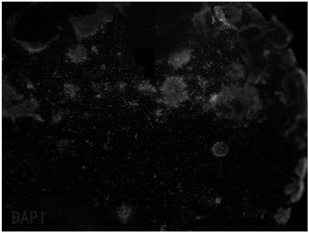
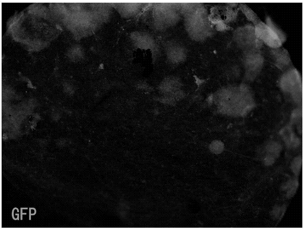
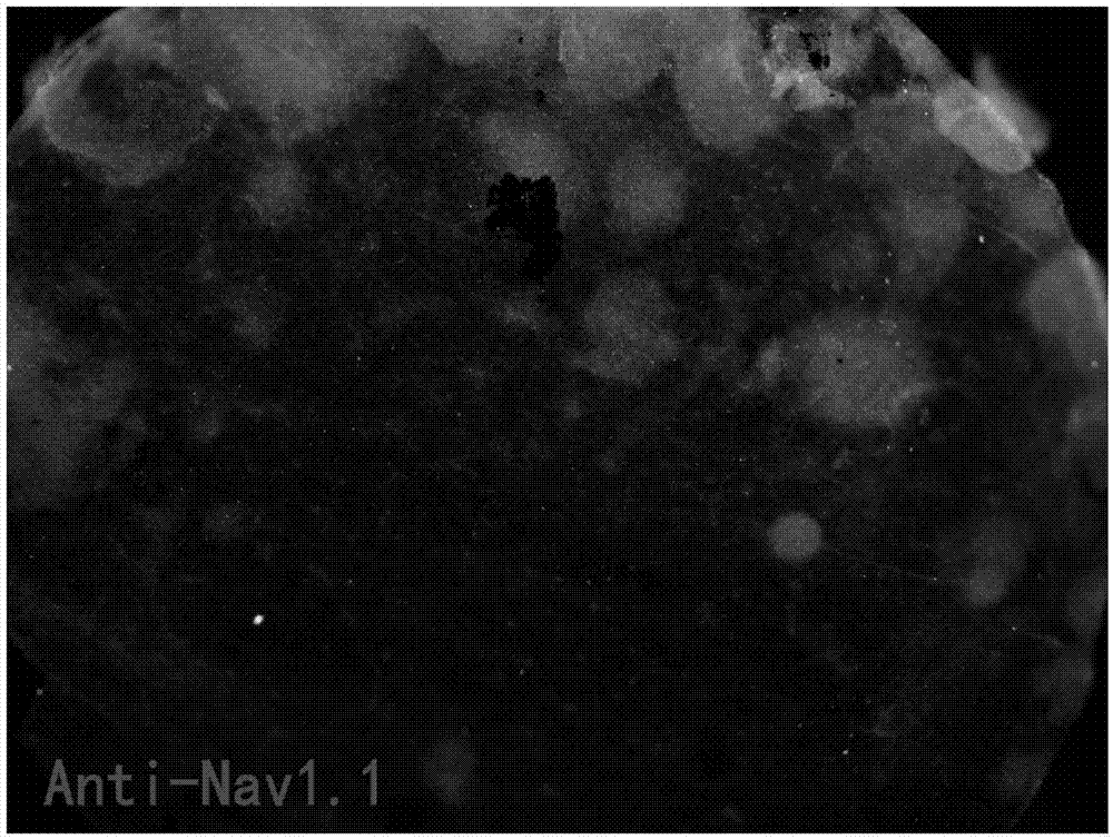
Embodiment 1
[0018] 1. Human iPSc-derived 3D retinal isolation and digestion:
[0019] 1. Using the method of mechanical separation, put the nerve fiber layer of human iPSc-derived 3D retina cultured for 50-60 days (specific preparation method reference: XiufengZhong, et al. Generation of three-dimensional retinal tissue with functional photoreceptors from human iPSCs, NatCommun.2014Jun10; 5:4047) Put it into D-Hanks solution, use a 0.45mm needle to separate the tissue under a 50 times upright microscope, discard the pigmented part (pigment layer) in the retinal tissue, and keep the golden yellow tissue part (nerve fiber layer); the separated nerve fiber Use a pipette to transfer the layered tissue to a 3.5cm culture dish, rinse with PBS for 10 minutes; absorb the PBS, digest the cells with accutase cell digestion solution, and place them in a 37°C incubator for 30 minutes; at the end of the process, the cells can be sucked into the centrifuge tube and blown. Under a 10x upright microscope...
PUM
 Abstract
Description
Claims
Application Information