GDNF-carrying microbubble preparation and method for making the same

A technology of microbubble and floating method, which is applied in the direction of pharmaceutical formulations, medical preparations containing active ingredients, peptide/protein components, etc., to achieve the effect of promoting blood-brain barrier penetration and increasing effective drug concentration

Active Publication Date: 2012-09-12
PEKING UNIV
View PDF5 Cites 12 Cited by
  • Summary
  • Abstract
  • Description
  • Claims
  • Application Information

AI Technical Summary

Problems solved by technology

However, at present, there is no report on the research on the treatment of various central nervous system diseases by using ...

Method used


Image

  • GDNF-carrying microbubble preparation and method for making the same
  • GDNF-carrying microbubble preparation and method for making the same
  • GDNF-carrying microbubble preparation and method for making the same

Examples


Embodiment 1

[0040] Example 1. Preparation of GDNF-loaded microbubble contrast agent and its research on opening the blood-brain barrier

[0041] 1) Preparation of GDNF-loaded microbubbles

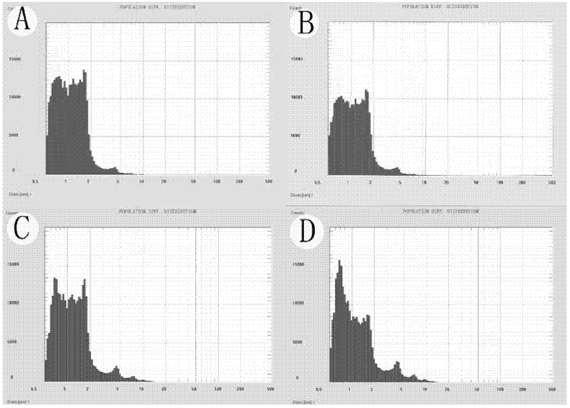
[0042] A certain amount of DSPC (distearoylphosphatidylethanolamine, 10.64mg), DSPE-PEG2k (polyethylene glycol-distearoylphosphatidylethanolamine, 2.10mg), DSPE-PEG2k-Biotin (polyethylene glycol) was added in a certain proportion. Ethylene glycol-distearoylphosphatidylethanolamine-biotin, 2.26 mg) was dissolved in 0.5 ml of chloroform and mixed on a vortex mixer. in dry N 2 Remove chloroform under the action of flow to form a uniform film of phospholipid on the test tube wall, and dry it in a vacuum oven for more than 2 hours. Then add the Tris buffer solution of 5ml degassed pH7.4 in the test tube that contains dry phospholipid film: containing glycerol (10% volume fraction) and propylene glycol (10% volume fraction) obtain the phospholipid solution of certain concentration. Heat the phospholipid s...

Embodiment 2

[0053] Example 2. Determining the optimal parameters of MRI-guided low-frequency focused ultrasound combined with targeted microbubbles for local opening of the BBB

[0054] Taking ultrasonic irradiation time, microbubble dose, delay time (interval between microbubble injection time and acoustic shock time), ultrasonic frequency, and sound pressure as influencing factors, three levels were selected for each factor, and an orthogonal experimental design was adopted (see Table 1). , taking Evans blue (EB, Evens blue, Sigma company, E2129) exudation as an indicator of the blood-brain barrier passage rate (EB exudation), to determine the local opening of the BBB by MRI-guided low-frequency focused ultrasound combined with targeted microbubbles, Optimal parameters for increasing central GDNF levels.

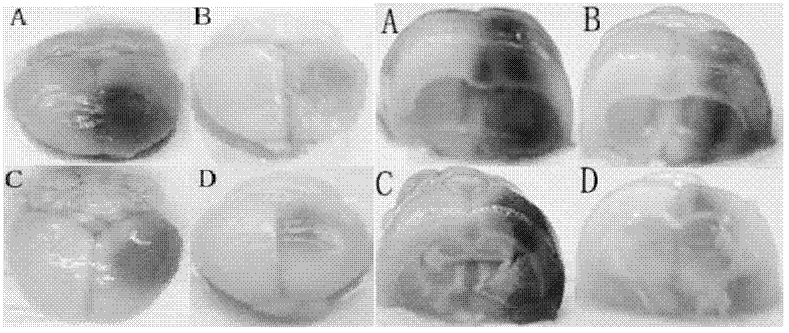
[0055] Test method: The permeability is measured by the quantitative estimation of the exudation of EB. Take the rat brain tissue perfused with PBS, immerse the partially blue-staine...

Embodiment 3

[0068] Embodiment 3, the mensuration of protein content in brain

[0069] The ultrasonic conditions of this embodiment are the optimal parameter conditions obtained in Example 2, that is, the probe frequency is 1 MHz, the amount of microbubbles is 0.5 ml, the irradiation time is 60 s, the sound pressure is 0.8 MPa, and the time delay is 60 s.

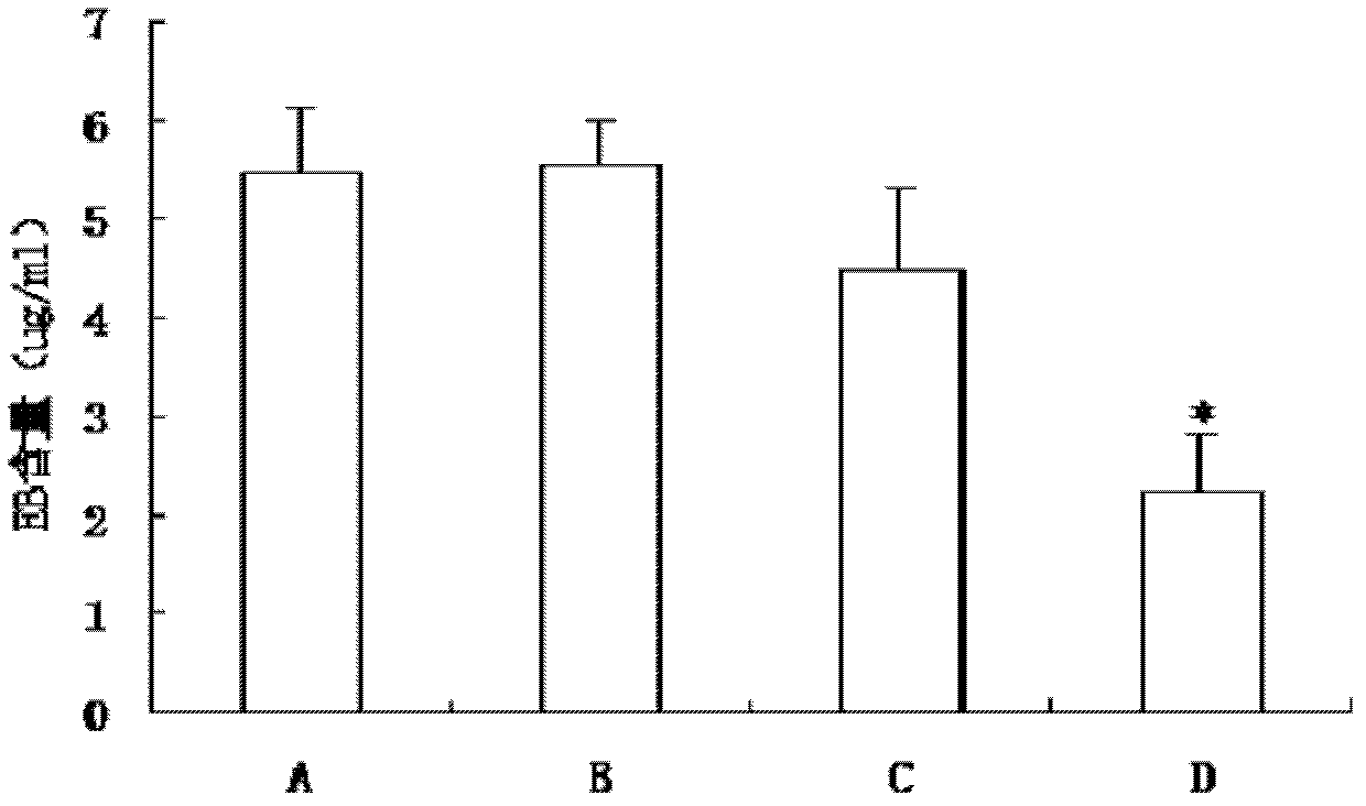
[0070] Ultrasound combined with microbubbles to open the blood-brain barrier, after intravenous injection of protein, the determination of protein content in the brain and the comparison of the two protein transport methods. Detect the amount of protein and the amount of protein entering the brain tissue after ultrasound combined with microbubbles to open the BBB.

[0071] (1) Due to the high price of GDNF, another low-priced alternative protein BSA was used to simulate the experimental conditions to determine the amount entering the brain tissue.

[0072] Divided into two groups: ultrasound + microbubbles + protein (group A); ultrasou...


PUM

PropertyMeasurementUnit
The average particle sizeaaaaaaaaaa

Abstract

The invention discloses a GDNF-carrying microbubble preparation and a method for making the same. The GDNF-carrying microbubble is composed of a lipid bilayer internally wrapping biological inert gas and biotinylated GDNF connected to the outside of the lipid bilayer. Employing MRI real-time guided low-frequency focused ultrasound combined with BBB opening by the GDNF-carrying microbubble to irradiate a craniocerebral parietal cortex area of a rat (the optimal parameters are set to be: probe frequency 1 Hz, microbubble 0.5 mL, irradiation period 60 s, sound pressure 0.8 MPa, and time-delay 60s) can promote GDNF to penetrate the BBB and increase the effective drug concentration of GDNF in the central nervous system. The pharmacological research after effective macromolecule breaking through of BBB by low-frequency focused ultrasound combined with target microbubble technique will further increase the advantage of neurotrophic factors in treatment of brain diseases and provide the scientific evidence for GDNF to treat nervous centralis diseases.

Description

technical field [0001] The invention relates to a GDNF-loaded microbubble preparation and a preparation method thereof. Background technique [0002] Glial cell line derived neurotrophic factor (GDNF) is one of the most representative members of the neurotrophic factor family. It was first discovered in the B49 glial cell line of rats. It is a 24kDa macromolecular protein. GDNF widely exists in the developing central nervous system and mature brain tissue, and is expressed at high levels in the striatum, thalamus, cortex and hippocampus, and has effects on various neurons such as glial cells, serotonergic neurons and dopamine Neurons have growth-promoting, protective and repairing functions, and can also regulate noradrenergic and GABAergic pathways. The role of GDNF in neurodegenerative diseases such as Parkinson's disease and Alzheimer's disease has been recognized. Recent clinical and animal experiments have shown that GDNF plays an important role in the treatment of de...

Claims


Application Information

Patent Timeline
IPC IPC(8): A61K9/127A61K38/19A61P25/16A61P25/24A61P25/28A61P25/30
Inventor 陈芸王峰石宇陆林孙成玉刘俐郑海荣
Owner PEKING UNIV
Why Eureka
Try Eureka