Angiography method
An angiography and sub-image technology, applied in the field of image processing in the medical field, can solve the problems of observation, long imaging time, unfavorable vascular imaging, etc., and achieve the effect of ensuring the imaging speed and improving the effect
- Summary
- Abstract
- Description
- Claims
- Application Information
AI Technical Summary
Problems solved by technology
Method used
Image

Examples
Embodiment 1
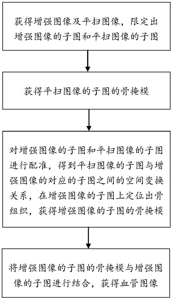
[0029] Example 1: Please refer to figure 1 As shown, the angiography method in the embodiment of the present invention specifically includes the following steps:
[0030] Step S1. Obtain an enhanced image and a plain-scan image, and define a sub-image of the enhanced image and a sub-image of the scanned image. Defining can be interpreted as obtaining by cutting and other means. The enhanced image and the unenhanced image may be scanned by X-ray imaging equipment, such as CT (Computed Tomography, computerized tomography) equipment, and the enhanced image may be a contrast image obtained by enhanced CT scanning. Enhanced CT scanning includes but is not limited to intravenous injection of iodine-containing contrast agent (contrast agent) followed by CT scanning to increase the density difference between lesion tissue and adjacent normal tissue, thereby improving the display rate of lesion. Increased density of lesion tissue is called enhancement or enhancement. The mechanism is...
Embodiment 2
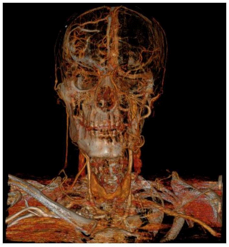
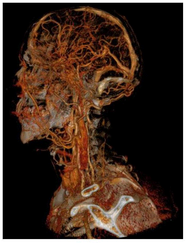
[0052] The angiography method of the present invention can also be applied to the cerebral angiography (DSA) or three-dimensional digital subtraction angiography (3DDSA) that utilizes the digital subtraction angiography machine to complete, and the difference between embodiment two and embodiment one mainly lies in: step S2 may also include combining the bone mask of the sub-image of the plain-scan image with the sub-image of the plain-scan image to obtain a sub-image of the plain-scan image with bone tissue removed.
[0053] Step S3 may also include combining the bone mask of the subimage of the enhanced image with the subimage of the enhanced image to obtain the subimage of the enhanced image with bone tissue removed; the subimage of the enhanced image with bone tissue removed subtracts the plain scan of the bone tissue Subgraphs of images to get subtracted images.
Embodiment 3
[0055] The angiographic method of the present invention can also be used to enhance MRA. The difference between the third embodiment and the first embodiment is that the bone mask is replaced with a mask of other tissues that need to be removed.
[0056] The angiography method of the above-mentioned embodiments of the present invention can be implemented in a computer-readable medium such as computer software, hardware or a combination of computer software and hardware. For hardware implementation, the embodiments described in this disclosure may be implemented on one or more Application Specific Integrated Circuits (ASICs), Digital Signal Processors (DSPs), Digital Signal Processing Devices (DAPDs), Programmable Logic Devices (PLDs) , field programmable gate array (FPGA), processor, controller, microcontroller, microprocessor, other electronic means for performing the functions described above, or a selected combination of the above means. In some cases, such embodiments may ...
PUM
 Abstract
Description
Claims
Application Information
 - R&D
- Intellectual Property
- Life Sciences
- Materials
- Tech Scout
- Unparalleled Data Quality
- Higher Quality Content
- 60% Fewer Hallucinations
Browse by: Latest US Patents, China's latest patents, Technical Efficacy Thesaurus, Application Domain, Technology Topic, Popular Technical Reports.
© 2025 PatSnap. All rights reserved.Legal|Privacy policy|Modern Slavery Act Transparency Statement|Sitemap|About US| Contact US: help@patsnap.com