Alpha 1-microglobulin detection kit
A detection kit and microglobulin technology, which is applied in the field of medical immunity, can solve the problems of reagent quality decline, poor reagent stability, and easy coalescence, etc., and achieve the effect of simple detection, low production cost and high sensitivity
- Summary
- Abstract
- Description
- Claims
- Application Information
AI Technical Summary
Problems solved by technology
Method used
Image

Examples
Embodiment 1
[0023] Example 1: Preparation of α1-microglobulin detection kit
[0024] The kit of the present invention relates to the main raw materials of reagents as follows:
[0025] 1. α1-microglobulin antibody: polyclonal antibody (commercially available).
[0026] 2. Latex: The present invention only exemplarily uses polystyrene latex particles (commercially available) with a diameter of 80-200 nm and carboxyl groups to carry out experiments.
[0027] The preparation of the main reagent of this embodiment is as follows:
[0028] Reagent R1: phosphate buffer solution containing 2.5% PEG6000 (polyethylene glycol 6000), 95mmol / L NaCl, this reagent is a colorless transparent solution.
[0029] Reagent R2: polystyrene latex particles with a particle diameter of 120 nm were sensitized with an anti-human α1-microglobulin antibody. The reagent is a milky white solution. Specific steps are as follows:
[0030] 1. Take 1ml (100mg / ml) latex, wash 3 times with 0.02M (mol / L), pH 5.0 MES solu...
Embodiment 2
[0037] Example 2: Determination of α1-microglobulin
[0038] Detection tool: Hitachi 7060 automatic analyzer.
[0039] Analysis method: two-point endpoint method; main wavelength: 600nm, secondary wavelength: -: sample volume: 2ul (microliter); R1: 200ul; R2: 50ul; reaction direction: rising; measurement temperature: 37 ° C; sample mixed with R1 After mixing, read the absorbance A1 at 10 seconds; add R2 at 3-5 minutes, and read the absorbance A2 after 5 minutes. The reaction absorbance was calculated as the difference between A2 and A1.
[0040] Calculation method: multi-point calibration, the dose / response curve is made according to the absorbance and reference serum value, and the sample content can be calculated on the dose / response curve according to its absorbance value.
Embodiment 3
[0041] Example 3 α1-microglobulin detection kit performance evaluation
[0042] 1. Analytical Sensitivity Assessment
[0043] Use 5% bovine serum albumin solution as a blank sample, and the blank sample should not contain the analyte. 20 consecutive detections were performed on the biochemical analyzer, and the mean and standard deviation SD of the 20 results were calculated. The detection limit of the reporting method is the mean value of the blank plus two standard deviations (+2SD). As can be seen from Table 1, the sensitivity is 1.03mg / L.
[0044] Table 1
[0045]
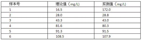
[0046] 2. High value linear evaluation
[0047] 1 part of low-value serum (5mg / L) and 1 part of high-value serum (120mg / L) were divided into 9:1, 4:1, 2:1, 1:2, 1:3, 1:9 (sample order No. 1 to No. 6), prepared 6 samples with different concentrations, each sample was repeatedly measured twice, as can be seen from Table 2, the highest detection range of the detection kit of the present invention can reach...
PUM
| Property | Measurement | Unit |
|---|---|---|
| particle size | aaaaa | aaaaa |
| diameter | aaaaa | aaaaa |
| Sensitivity | aaaaa | aaaaa |
Abstract
Description
Claims
Application Information
 - R&D
- Intellectual Property
- Life Sciences
- Materials
- Tech Scout
- Unparalleled Data Quality
- Higher Quality Content
- 60% Fewer Hallucinations
Browse by: Latest US Patents, China's latest patents, Technical Efficacy Thesaurus, Application Domain, Technology Topic, Popular Technical Reports.
© 2025 PatSnap. All rights reserved.Legal|Privacy policy|Modern Slavery Act Transparency Statement|Sitemap|About US| Contact US: help@patsnap.com