Human pancreatic cancer nude mouse model construction method and use thereof

A construction method and pancreatic cancer technology, applied in the field of construction of human pancreatic cancer nude mouse model, can solve the problems of high tumor formation rate, low cell tumor formation rate, and long tumor formation incubation period, and achieve high tumor formation rate, tumor formation fast effect

Active Publication Date: 2016-08-10
ZHEJIANG CHINESE MEDICAL UNIVERSITY
View PDF1 Cites 6 Cited by
  • Summary
  • Abstract
  • Description
  • Claims
  • Application Information

AI Technical Summary

Problems solved by technology

[0005] The purpose of the present invention is to establish an animal model of pancreatic cancer suitable for in vivo imaging research. The model has a short incubation period for tumor formation, a high tumor formation rate, tumor growth in line with the characteristics of pancreatic cancer, and has stable, high-efficiency, and good linear bioluminescence characteristics. It overcomes the shortcomings of low tumor formation rate, long incubation period of tumor formation and abnormal tumor growth of bioluminescent labeled cells constructed by lentiviral vectors, and is especially suitable for in vivo imaging research of pancreatic cancer

Method used


Image

  • Human pancreatic cancer nude mouse model construction method and use thereof
  • Human pancreatic cancer nude mouse model construction method and use thereof
  • Human pancreatic cancer nude mouse model construction method and use thereof

Examples


Embodiment 1

[0033] Embodiment 1: the determination of puromycin optimal screening concentration

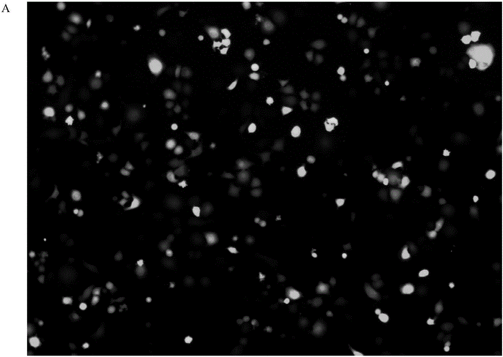
[0034] (1) Materials: puromycin, human pancreatic cancer PANC-1 cells (purchased from ATCC, USA).

[0035] (2) Experimental method: Digest the PANC-1 cells in the logarithmic growth phase with 0.25% trypsin, inoculate them in a 6-well plate at a cell density of 50%, add DMEM containing 10% fetal bovine serum to culture in high glucose Incubate overnight at 37°C in a 5% CO2 incubator. On the second day, different concentrations of puromycin (1 μg / ml, 2.5 μg / ml, 5 μg / ml, 7.5 μg / ml, 10 μg / ml) were added, and new media containing different concentrations of puromycin were replaced every 2 days. The minimum concentration at which all cells died within 4 days was selected as the screening concentration.

[0036] (3) Experimental results: On the second day of culture, a large number of cells died in the wells with a dose of 5 μg / ml or more of puromycin. After 3 days of culture, different degrees ...

Embodiment 2

[0037]Example 2: Construction of PANC-1 cells stably expressing luciferase

[0038] (1) Materials: plasmid vector GV258 with luciferase gene, puromycin, Nucleofector TM Solution SE Kit.

[0039] (2) Experimental method

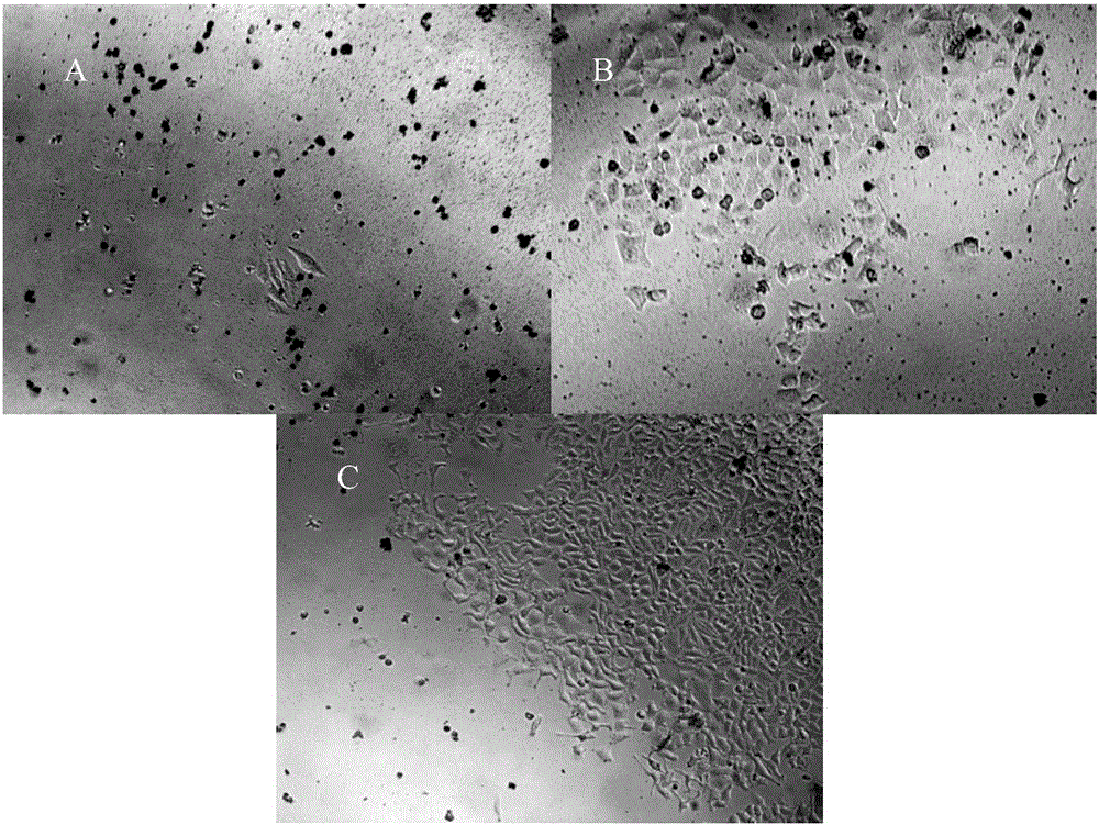
[0040] 1.1 Cell transfection

[0041] Take the 4th passage PANC-1 cells after recovery, digest the cells with 0.25% trypsin+0.02% EDTA, collect the cells by centrifugation at 1000r / min for 5min, take 5×10 6 cells, flick the bottom of the tube to mix the cells to Resuspend the cells in SE Solution, add 8 μg of the GV258 plasmid vector expressing luciferase, gently add the cell suspension to the electroporation cup, 4D-Nucleofector TM System nucleofection system for cell transfection with DN-100 program. Transfer the cells in the electroporation cup to a 6-well cell culture plate with 37°C preheated medium, 37°C, 5% CO 2 Cultivate in the incubator for 48h.

[0042] 1.2 Screening of PANC-1 cells stably expressing luciferase

[0043] Collect the PANC-1 ...

Embodiment 3

[0046] Example 3: In vivo imaging system detects the expression of luciferase in PANC-1-LUC cells

[0047] (1) Materials: PANC-1-LUC cells, D-fluorescein potassium salt, isoflurane.

[0048] Experimental animals: SPF grade BALB / c-nu / nu nude mice, male, 4-5 weeks old.

[0049] (2) Experimental method

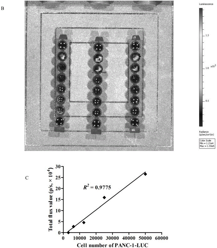
[0050] 1.1 In vitro bioluminescence detection

[0051] Digest and count the PANC-1-LUC cells in the logarithmic growth phase, and adjust the cell concentration to 5×10 5 cells / ml, 100 μl per well was inoculated in a black 96-well plate, and the cells were sequentially diluted one by one. Three parallel wells were set up for each order of magnitude of cells, and 100 μl of D-luciferin (150 μg / ml) was added to each well. detected in the imaging system.

[0052] 1.2 In vivo bioluminescence detection

[0053] Digest and count the PANC-1-LUC cells in the logarithmic growth phase, and adjust the cell density to 5×10 7 cells / ml, and then diluted to 2.5×10 7 pcs / ml, 1×10 7 pcs / ml,...


PUM


Abstract

The invention relates to a novel human pancreatic cancer nude mouse model construction method for living imaging research and a use thereof. A human pancreatic cancer PANC-1 cell is stably transfected with a plasmid carrying a luc gene through a Lonza nuclear transfection system, a cell strain PANC-1-LUC for stable luciferase expression is screened through puromycin and PANC-1-LUC nude mouse transplant subcutaneous sarcoma is further constructed. An in vitro and vivo bioluminescence detection result proves that the PANC-1-LUC cell strain can stably express luciferase for a long time. Compared with a lentiviral vector-mediated bioluminescence pancreatic cancer nude mouse model, the built pancreatic cancer nude mouse model has a short tumor formation incubation period and a high tumor formation rate, satisfies PANC-1 transplantation tumor growth features and is suitable for living imaging research.

Description

technical field [0001] The invention relates to a new construction method and application of a nude mouse model of human pancreatic cancer suitable for live imaging research. Background technique [0002] Pancreatic cancer is a common malignant tumor of the digestive tract worldwide, and it is one of the malignant tumors with the worst prognosis. The low early diagnosis rate and postoperative metastasis are the main reasons for the high mortality rate of pancreatic cancer. It is difficult to obtain fresh pathological tissue samples of pancreatic cancer at different stages in clinical practice, which greatly restricts the research on the occurrence and development of pancreatic cancer. In recent years, in vivo imaging technology has been widely used in tumor research as an efficient and sensitive new detection method, especially bioluminescent imaging technology, which has the advantages of small signal interference, low imaging background, high signal-to-noise ratio and tiss...

Claims


Application Information

Patent Timeline
Patent Type & Authority Applications(China)
IPC IPC(8): C12N15/85C12N15/53A01K67/027
CPCA01K67/0271A01K2207/12A01K2227/105A01K2267/0331C12N9/0069C12N15/8509C12N2800/107
Inventor 屠珏陈民利凌云黄宇齐月寒
Owner ZHEJIANG CHINESE MEDICAL UNIVERSITY
Why Eureka
Try Eureka