Cysteine engineered fibronectin type III domain binding molecules

A cysteine, fibronectin technology, applied in the direction of peptide/protein components, immunoglobulins, animal/human proteins, etc.

Active Publication Date: 2016-08-24
JANSSEN BIOTECH INC
View PDF22 Cites 5 Cited by
  • Summary
  • Abstract
  • Description
  • Claims
  • Application Information

AI Technical Summary

Problems solved by technology

However, modification in cysteine ​​thiol groups obtained by mutation of various amino acid residues of the protein to cysteine ​​amino acids can be problematic, especially at unpaired (free Cys) residues or relative In the case of those residues susceptible to reaction or oxidation

Method used


Image

  • Cysteine engineered fibronectin type III domain binding molecules
  • Cysteine engineered fibronectin type III domain binding molecules
  • Cysteine engineered fibronectin type III domain binding molecules

Examples


example 1

[0364] Example 1: Construction of Tencon library

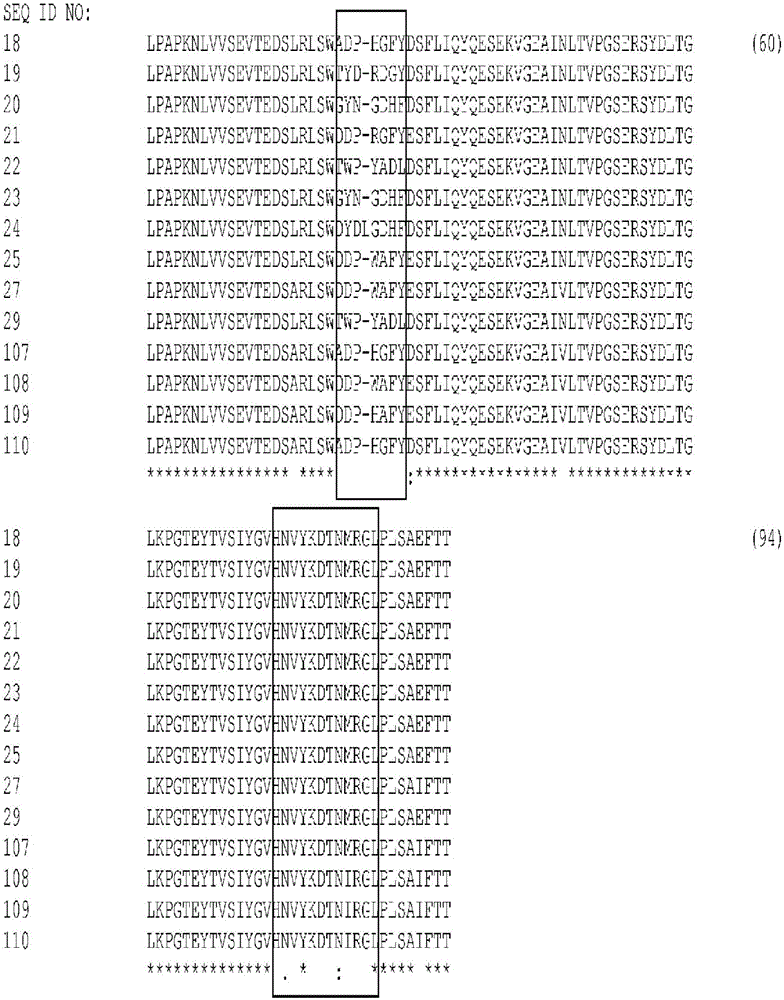
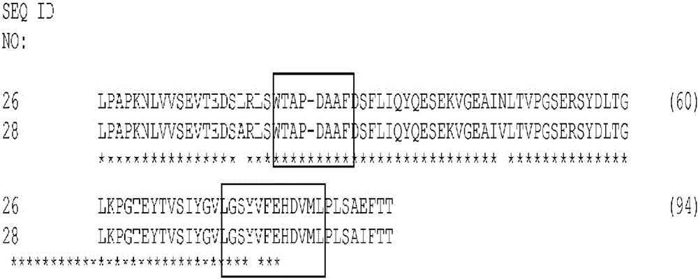
[0365] Tencon (SEQ ID NO: 1) is an immunoglobulin-like scaffold designed from the consensus sequence of fifteen FN3 domains from human tenascin-C fibronectin (FN3) domain type III (Jacobs et al., Protein Engineering, Design, and Selection, 25:107-117, 2012; US Patent Publication 2010 / 0216708). The crystal structure of Tencon shows six surface-exposed loops linking seven β-strands. Selected residues within the or each loop can be randomized in order to construct a type III fibronectin (FN3) domain library that can be used to select novel molecules that bind specific targets.

[0366] Tencon:

[0367] LPAPKNLVVSEVTEDSLRLSWTAPDAAFDSFLIQYQESEKVGEAINLTVPGSERSYDLTGLKPGTEYTVSIYGVKGGHRSNPLSAEFTT (SEQ ID NO 1):

[0368] Construction of TCL1 library

[0369] A library TCL1 designed to randomize only the FG loop of Tencon (SEQ ID NO: 1) was constructed for use with the cis-display system (Jacobs et al., Protein Engineering, Desig...

example 2

[0391] Example 2: Selection of Type III Fibronectin (FN3) Domains that Bind EGFR and Inhibit EGF Binding

[0392] library screening

[0393] Cis display was used to select EGFR binding domains from TCL1 and TCL2 libraries. The recombinant human extracellular domain of EGFR fused to IgGl Fc (R&D Systems) was biotinylated using standard methods and used for panning (residues 25-645 of full-length EGFR of SEQ ID NO: 73). For in vitro transcription and translation (ITT), 2-6 μg of library DNA was incubated with 0.1 mM intact amino acids, 1X S30 master mix components and 30 μL S30 extract (Promega) in a total volume of 100 μL and incubated at 30°C . After 1 hour, 450 μL of blocking solution (PBS pH 7.4 supplemented with 2% bovine serum albumin, 100 μg / mL herring sperm DNA and 1 mg / mL heparin) was added and the reaction was incubated on ice for 15 minutes. EGFR-Fc was assembled at 1:1 and 10:1 molar ratios of EGFR to EGF by mixing recombinant human EGF (R&D Systems) and bioti...

example 3

[0411] Example 3: Characterization of EGFR-binding FN3 domains that inhibit EGF binding

[0412] Large scale expression, purification and endotoxin removal

[0413] The nine FN3 domains shown in Table 4 were enlarged to provide more material for detailed characterization. Using overnight cultures containing each EGFR-binding FN3 domain variant, dilute the overnight culture 1 / 80 with 0.8 L of Terrific broth medium supplemented with 100 μg / mL ampicillin and inoculate into fresh medium, Incubate with shaking at 37°C. When the optical density at 600 nm reached about 1.2-1.5 by adding IPTG to a final concentration of 1 mM, the culture was induced and the temperature was lowered to 30°C. After 4 hours, cells were collected by centrifugation and the cell pellet was stored at -80°C until required for use.

[0414] For cell lysis, dissolve the thawed pellet at 5 mL per gram of pellet The ratio was resuspended in supplemented with 25U / mL (Sigma-Aldrich) and 1kU / mL rLysozyme ...


PUM

PropertyMeasurementUnit
affinityaaaaaaaaaa
sizeaaaaaaaaaa

Abstract

The invention discloses cysteine engineered monospecific and bispecific EGFR and / or c-Met FN 3 domain containing molecules comprising one or more free cysteine amino acids. The molecules are prepared by mutagenizig a nucleic acid sequence of a parent molecule and replacing one or more amino acid residues by cysteine to encode the cysteine engineered FN3 domain containing monospecific or bispecific molecules; expressing the cysteine engineered FN3 domain containing molecules: and recovering the cysteine engineered FN3 domain containing molecule. Isolated cysteine engineered monospecific or bispecific FN3 domain containing molecules may be covalently attached to a detection label or a drug moiety and used therapeutically.

Description

technical field [0001] The present invention relates to binding molecules engineered with cysteine ​​residues, as well as methods of making them and using them. More specifically, the present invention relates to fibronectin type III (FN3) domain molecules that bind to cysteine ​​engineered EGFR and / or c-Met. Background technique [0002] Epidermal growth factor receptor (EGFR or ErbB1 or HER1) is a 170kDa transmembrane glycoprotein encoded by the c-erbB1 proto-oncogene. EGFR is a member of the human epidermal growth factor receptor (HER) family of receptor tyrosine kinases (RTKs), which includes HER2 (ErbB2), HER3 (ErbB3) and HER4 (ErbB4). These RTKs share a homologous structure consisting of a ligand-binding extracellular domain (ECD), a single transmembrane domain, and an intracellular domain containing a catalytic kinase domain and a C-terminal tail. EGFR signaling is initiated by ligand binding, which then induces conformational changes, dimerization, and trans-autop...

Claims


Application Information

Patent Timeline
Patent Type & Authority Applications(China)
IPC IPC(8): A61K38/22C12N1/20
CPCA61K2039/505A61P35/00A61P43/00C07K14/78C07K16/2863C07K2317/31C07K2317/73C07K2317/76C07K2317/92C07K2318/20C07K2319/00C07K2319/30C07K2319/31G01N33/74C12N15/70
Inventor S.戈德伯格S.贾科布斯T.林K.奥内尔
Owner JANSSEN BIOTECH INC
Why Eureka
Try Eureka