
Method and kit for detecting free rare tumor cells in human biofluid sample

A technology of biological liquid and tumor cells, applied in the field of biological and medical detection, can solve the problems of loss of CTC, inability to accurately identify tumor cells, low expression of epithelial markers, etc., and achieve the effect of reducing the loss of CTC

Active Publication Date: 2016-09-21
上海乾翼生物科技有限公司
View PDF5 Cites 20 Cited by
  • Summary
  • Abstract
  • Description
  • Claims
  • Application Information

AI Technical Summary

Problems solved by technology

[0004] However, the CTC detection method represented by the CellSearch system has several defects.
First, the enrichment method of the CellSearch system is not sensitive, the detection rate is low, and a considerable part of the CTC is lost during the enrichment process
In fact, the enrichment of CTCs is not only cumbersome and complicated, but also will inevitably lose varying amounts of CTCs during the enrichment process.
Second, the tumor cell identification method based on EpCAM+ / DAPI+ / CK+ / CD45- is not strictly accurate, it does not use any tumor-specific markers, and actually identifies cells of epithelial origin rather than tumor cells
The latest research shows that cells of epithelial origin can be detected in the blood of patients with benign diseases and even healthy people, so there is a possibility of false positives in this identification method
At the same time, studies have found that the epithelial-mesenchymal transition (EMT) of tumor cells during metastasis will lead to no or low expression of epithelial markers in CTCs, so they may be missed
Third, since the above-mentioned CTC identification method based on immunofluorescence staining uses fixation and nuclear staining, it is difficult for CTCs stained by immunofluorescence to be further used for sequencing analysis and in vitro culture, and sequencing is currently the most important method for tumors. Molecular detection means
[0005] Almost all current CTC detection methods have similar defects to the CellSearch system, the enrichment steps are cumbersome and complex, and CTCs are lost, while the CTC identification method based on immunofluorescence staining cannot accurately identify tumor cells, and limits subsequent gene sequencing analysis and in vitro culture carry out

Method used


Image

  • Method and kit for detecting free rare tumor cells in human biofluid sample
  • Method and kit for detecting free rare tumor cells in human biofluid sample
  • Method and kit for detecting free rare tumor cells in human biofluid sample

Examples


Embodiment 1

[0094] Glucose analogue uptake assay in tumor cells

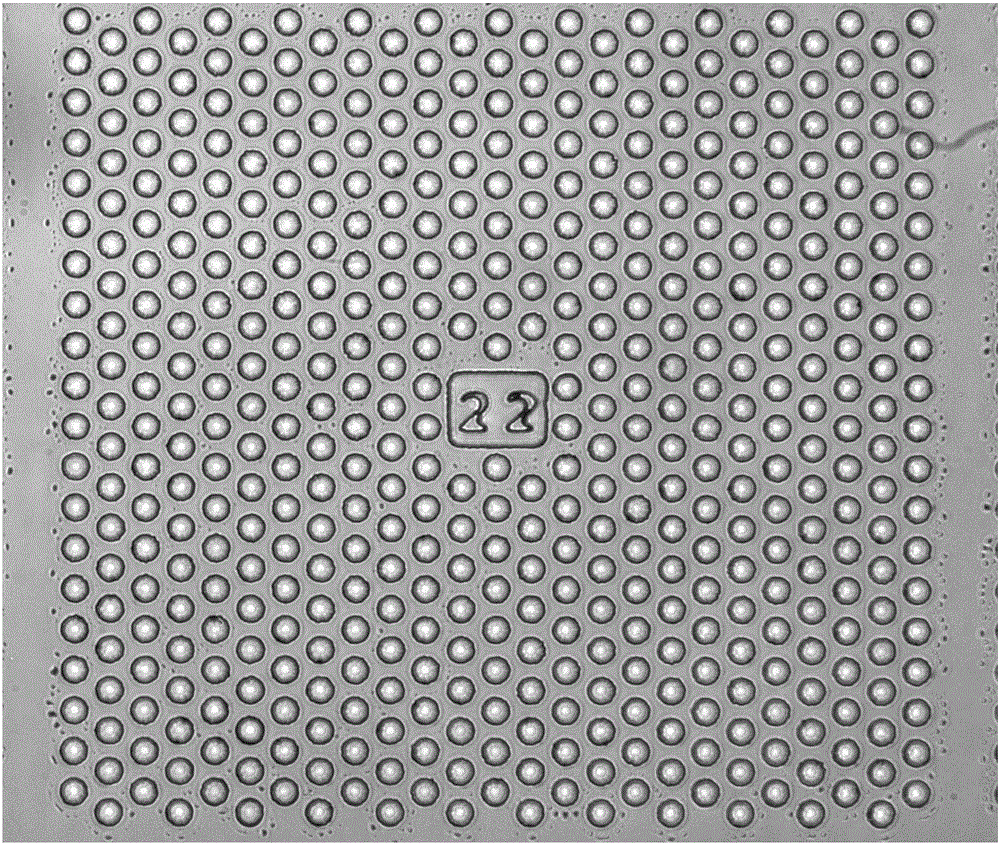
[0095] Provide a microwell array PDMS chip, its structure is as follows figure 1 As shown, each microwell is 20 microns in diameter and 30 microns deep, and every 500 microwells form a small area and are marked with numbers to realize the addressability of each microwell. A typical microwell chip consists of 400 such as Figure 1 The numerically coded small regions shown consist of 200,000 microwells.

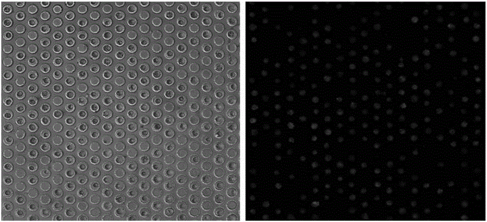
[0096] Add the cell suspension of lung cancer cell H1650 on the microwell array chip, wash it with glucose-free cell culture medium RPMI-1640 after standing, as in figure 2 Cells are shown in most of the wells and at most one cell per well, with cells in the center of the well.

[0097] The tumor cells in the microwells were starved by incubating the cells with glucose-free cell culture medium RPMI-1640 for 10 minutes. Then, cell culture medium RPMI-1640 containing 2-NBDG (0.3 mM concentration) was added to incubate the...

Embodiment 2

[0099] Detection of Tumor Cells in Pleural Effusion Samples of Lung Cancer Patients

[0100] In this embodiment, the method includes the following steps:

[0101](1) After filtering 40 milliliters of pleural effusion of a lung cancer patient with 150 mesh gauze, centrifuge (500g, 5 minutes) to separate the cells, add 5 milliliters of red blood cell lysate (BD Company) to lyse in the dark for 5 minutes, and centrifuge again (500g, 5 minutes) ), after discarding the supernatant, resuspend and wash the cells with Hank's Balanced Salt Solution (HBSS), centrifuge (500g, 5 minutes), discard the supernatant, add 2 milliliters of HBSS to resuspend the cells;

[0102] (2) After cell counting, take 500 microliters of cell suspension (about 1 million cells), add 2 microliters of APC-labeled CD45 antibody, and incubate for 1 hour on an invertor;

[0103] (3) Centrifuge, discard the supernatant and dilute the cells with HBSS, and drop the cell suspension on 2 microwell array chips (each c...

Embodiment 3

[0113] Detection of glucose analog uptake by tumor cells in peripheral blood samples of lung cancer patients

[0114] In this embodiment, the method includes the following steps:

[0115] (1) Take 1 ml of peripheral blood samples from patients with lung cancer, first centrifuge at low speed (200g, 5 minutes) to remove the platelet-rich plasma in the upper layer, resuspend the remaining cells in HBSS, add erythrocyte lysate (BD company) to lyse in the dark for 5 minutes, and again Centrifuge (500g, 5 minutes), discard the supernatant, resuspend and wash the cells with Hank's Balanced Salt Solution (HBSS), centrifuge (500g, 5 minutes), discard the supernatant, add 2 ml of HBSS to resuspend the cells;

[0116] (2) After counting cells, add CD45 antibody-labeled magnetic balls (Stemcell) according to the cell number 1:20, invert and incubate on an invertor, add 4 microliters of APC-labeled CD45 antibody after 15 minutes and continue inverting for 45 minutes;

[0117] (3) Transfer...


PUM

PropertyMeasurementUnit
Diameteraaaaaaaaaa

Abstract

The invention provides a method and a kit for detecting free rare tumor cells in a human biofluid sample, and concretely provides a method for identifying active tumor cells through detecting the glucose intake ability of karyotes in the biofluid sample of a tumor patient and the expression of a leucocyte marker CD45. The method and the kit have the advantages of simple and rapid operation, no need of preconcentration of the tumor cells in the sample, and realization of extremely small loss of the tumor cells; and the result obtained through using the metabolism function-based identification method for identifying the free tumor cells in the fluid sample is more accurate than results obtained through common immunofluorescence dyeing methods.

Description

technical field [0001] The invention relates to the field of biological and medical detection. More specifically, the present invention relates to a detection method and a kit for free rare tumor cells in human peripheral blood or pleural effusion samples. The present invention provides a method and kit for rapidly detecting active rare tumor cells from human biological fluid samples (such as blood, pleural effusion, pericardial effusion, preferably peripheral blood or pleural effusion samples of tumor patients). Background technique [0002] Circulating Tumor Cells (CTCs) in human peripheral blood refer to tumor cells that shed from tumor lesions and enter the peripheral blood circulation. It represents the molecular characteristics of tumor lesions and can develop into tumor metastatic lesions under certain conditions. Therefore, the counting, molecular detection and in vitro culture of CTCs in blood have attracted more and more attention. However, CTCs are extremely rar...

Claims


Application Information

Patent Timeline
IPC IPC(8): G01N21/64
CPCG01N21/6486
Inventor 施奇惠陆舜邓宇亮汤寅
Owner 上海乾翼生物科技有限公司
Learn More