
Magnetic particle-based quantitative chemiluminescent assay kit for anti-ribosome P protein antibody IgG, and preparation and detection methods thereof

An anti-ribosome, magnetic particle technology, applied in chemiluminescence/bioluminescence, analysis by chemical reaction of materials, measurement devices, etc., can solve problems such as unseen

Inactive Publication Date: 2016-09-21
北京贝尔医疗设备有限公司
View PDF6 Cites 17 Cited by
  • Summary
  • Abstract
  • Description
  • Claims
  • Application Information

AI Technical Summary

Problems solved by technology

At present, the application of magnetic particle chemiluminescence analysis in anti-ribosomal P protein antibody IgG immunoassay products has not yet been seen.

Method used


Image

  • Magnetic particle-based quantitative chemiluminescent assay kit for anti-ribosome P protein antibody IgG, and preparation and detection methods thereof
  • Magnetic particle-based quantitative chemiluminescent assay kit for anti-ribosome P protein antibody IgG, and preparation and detection methods thereof
  • Magnetic particle-based quantitative chemiluminescent assay kit for anti-ribosome P protein antibody IgG, and preparation and detection methods thereof

Examples


Embodiment 1

[0067] Preparation of anti-ribosomal P protein antibody IgG calibrator:

[0068] a. Prepare anti-ribosomal P protein antibody IgG calibrator dilution:

[0069] Add 800ml of purified water, 11.2g of Tris, 8.6g of sodium chloride and 2ml of Proclin300 into the container, stir well until completely dissolved; use 4M HCL to adjust the pH of the solution to 7.0-7.5; add 40g of bovine serum albumin Add to the container, stir well until completely dissolved; then use 4M HCL to adjust the pH of the solution to 7.0-7.5; use purified water to dilute the solution to 1L, filter with a 0.2μm filter to obtain the anti-ribosomal P protein antibody IgG calibration Product diluent, store at 2-8°C until use;

[0070] b. Prepare anti-ribosomal P protein antibody IgG calibrator:

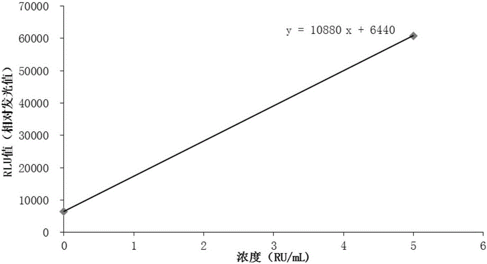
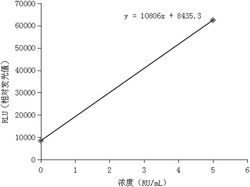
[0071] Dilute anti-ribosomal P protein antibody IgG with anti-ribosomal P protein antibody IgG calibrator diluent to each concentration point of 0, 5, 20, 50, 100, 200 RU / mL.

Embodiment 2

[0073] Preparation of anti-ribosomal P protein antibody IgG quality control:

[0074] Dilute the anti-ribosomal P protein antibody IgG with the above-mentioned anti-ribosomal P protein antibody IgG calibrator diluent to 20, 100 RU / mL at each concentration point.

Embodiment 3

[0076] Preparation of Reagent No. 1:

[0077] a. Prepare reagent No. 1 dilution:

[0078] Add 800ml of purified water, 12.1g of Tris, 5.8g of sodium chloride and 2ml of Proclin300 into the container, stir well until completely dissolved; add 5g of bovine serum albumin into the container, stir well until completely dissolved; dissolve the solution with 4M HCL Adjust the pH value of the solution to 7.0-7.5; dilute the solution to 1L with purified water, filter it with a 0.2μm filter to obtain the No. 1 dilution of the reagent, and store it at 2-8°C for later use;

[0079] b. Prepare reagent No. 1:

[0080] Dissolve the Rib-p antigen in purified water, dialyze it with a carbonate buffer solution with a concentration of 0.2M and a pH of 9.0 for 2 hours at 2-8°C, and then concentrate it to an antigen solution with a concentration of 2-4 mg / mL. Prepare a biotin solution with a concentration of 0.5-1.0 mg / ml in a carbonate buffer solution with a pH of 0.2M and a pH of 8.5-9; prepar...


PUM


Abstract

The invention discloses a magnetic particle chemiluminescence quantitative assay kit for anti-ribosomal P protein antibody IgG. The kit includes: anti-ribosomal P protein antibody IgG calibrator; Ribosomal P protein antigen and bovine serum albumin-labeled Tris buffer; alkaline phosphatase-labeled goat anti-human polyclonal antibody and bovine serum albumin in Tris buffer; streptavidin-labeled magnetic particles and bovine serum albumin Serum albumin in Tris buffer; wash solution. Based on the traditional membrane strip immunoassay and enzyme-linked immunosorbent assay, the detection method of the kit increases the sensitivity and linear range by 3-5 orders of magnitude, and realizes quantitative detection in a real sense, with rapid response, reliable results, and It can be used in conjunction with a fully automatic chemiluminescence immunoassay analyzer to realize fully automatic use, and has irreplaceable important value for clinical diagnosis.

Description

technical field [0001] The invention relates to the technical field of biological detection. More specifically, it relates to a magnetic particle chemiluminescence quantitative assay kit for anti-ribosomal P protein antibody IgG and a preparation and detection method. Background technique [0002] Anti-ribosomal P protein antibodies act directly on specific ribosomal phosphorylated proteins. Anti-Rib-P IgG is a highly specific indicator for the diagnosis of systemic lupus erythematosus (SLE), and it is almost undetectable in other autoimmune diseases. The positive rate of Anti-Rib-P IgG in SLE patients is between 5% ~46%. [0003] At present, there are membrane strip immunoassay and enzyme-linked immunosorbent assay for clinical detection of anti-ribosomal P protein antibody IgG. Membrane immunoassay uses membrane strip chromogenic technology, which is characterized by fixing several items in the same membrane strip for measurement, generally through manual or semi-automa...

Claims


Application Information

Patent Timeline
IPC IPC(8): G01N21/76G01N33/532G01N33/535
CPCG01N21/76G01N33/532G01N33/535
Inventor 不公告发明人
Owner 北京贝尔医疗设备有限公司
Learn More