Immune lateral chromatographic detection system as well as preparation method and application thereof
A technology of immune lateral chromatography and detection system, which is applied in the field of immune lateral chromatography detection system and its preparation, can solve the problems that the sensitivity, precision and accuracy of the detection and detection system cannot meet the existing needs, and achieve the extension of Effects of distance, reagent consumption reduction, and sensitivity improvement
- Summary
- Abstract
- Description
- Claims
- Application Information
AI Technical Summary
Problems solved by technology
Method used
Image
Examples
Embodiment 1
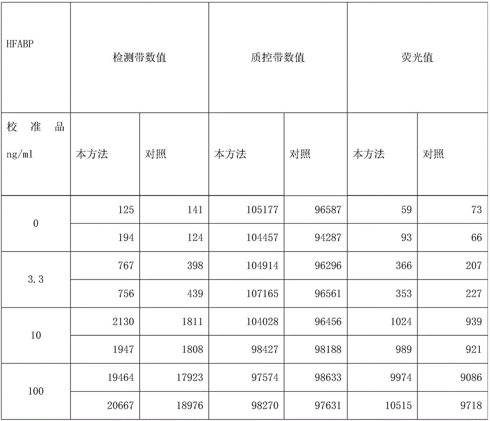
[0034] Example 1 (no marker pad, marker in test tube, with sample diluent)
[0035] Coating antibody: Dilute human heart fatty acid binding protein (HFABP) detection antibody 1 with coating buffer to a fixed concentration (2.0mg / ml), and control reagent 1 (goat anti-chicken IgY) to a fixed concentration (2.0mg / ml ), using BioDot’s XYZ3060 to coat the above two liquids on Sartorius nitrocellulose membrane 140 (NC membrane), wherein the quality control zone is located between the detection zone and the sample hole, and dried in a 37°C oven for 4 hours ,spare. Coating buffer is 0.01Mol / l phosphate buffer solution (PBs) plus 3% sucrose as protective agent.
[0036] Labeled antibodies: human heart fatty acid binding protein (HFABP) detection antibody 2 and control reagent 2 (chicken IgY) were fluorescently labeled with latex, stored in storage solution, and ready for use (50mMol / l Tris, 0.5%BSA, pH 7.8).
[0037] Marker preparation: Spray 5 microliters of the above-mentioned labe...
Embodiment 2
[0046] Example 2 (no marker pad, marker in tip, with sample diluent)
[0047] Coating antibody: Dilute C-reactive protein (CRP) detection antibody 1 with coating buffer to a fixed concentration (0.5mg / ml), and control reagent 1 (goat anti-chicken IgY) to a fixed concentration (2.0mg / ml), The above two liquids were coated on the Sartorius nitrocellulose membrane (NC) using XYZ3060 from BioDot Company, where the quality control zone was located between the detection zone and the sample hole, and dried in a 37°C oven for 4 hours before use. Coating buffer is 0.01Mol / l phosphate buffer solution (PBs) plus 3% sucrose as protective agent.
[0048] Labeled antibody: C-reactive protein (CRP) detection antibody 2 and control reagent 2 (chicken IgY) were fluorescently labeled with latex, stored in storage solution, and used for later use (50mMol / l Tris, 0.5% BSA, pH 7.8).
[0049] Marker preparation: Spray 5 microliters of the above-mentioned labeled antibody into the inner wall of the...
Embodiment 3
[0059] Example 3. (there is a marker pad, the coating reagent is an antigen, and there is no sample diluent)
[0060] Coating antibody: HCV (hepatitis C) detection antigen 1 was diluted to a fixed concentration (3.0mg / ml) with coating buffer, and the control reagent 1 (streptavidin SA) was diluted to a fixed concentration (2.0mg / ml ), using BioDot’s XYZ3060 to coat the above two liquids on a nitrocellulose membrane (NC membrane), wherein the quality control zone is located between the detection zone and the sample hole, and dried in a 37° C. oven for 4 hours. Coating buffer is 0.01Mol / l phosphate buffer solution (PBs) plus 3% sucrose as protective agent.
[0061] Labeled antibody: HCV (hepatitis C) detection antigen 2 and control antibody 2 (BSA-Biotin) were labeled with colloidal gold and stored in gold-labeled storage solution (50mMol / l Tris, 0.5% BSA, pH 7.8),
[0062] Marker pad preparation: Soak or spray the above-mentioned labeled antibody in or on the gold label pad ac...
PUM
 Abstract
Description
Claims
Application Information
 - R&D
- Intellectual Property
- Life Sciences
- Materials
- Tech Scout
- Unparalleled Data Quality
- Higher Quality Content
- 60% Fewer Hallucinations
Browse by: Latest US Patents, China's latest patents, Technical Efficacy Thesaurus, Application Domain, Technology Topic, Popular Technical Reports.
© 2025 PatSnap. All rights reserved.Legal|Privacy policy|Modern Slavery Act Transparency Statement|Sitemap|About US| Contact US: help@patsnap.com


