3D (three-dimensional) printing preparation method of orthopaedic external fixing support

A 3D printing and external fixation technology, applied in fractures, medical science, additive processing, etc., can solve the problems of high cost, low degree of fit, long customization time, etc., and achieve the effect of correcting mechanical strength and high degree of fit

Inactive Publication Date: 2016-12-14
QINGDAO UNIQUE PRODS DEV
View PDF8 Cites 21 Cited by
  • Summary
  • Abstract
  • Description
  • Claims
  • Application Information

AI Technical Summary

Problems solved by technology

[0003] The invention provides a 3D printing preparation method for orthopedic external fixation braces, which solves the problems of low degree of coincidence between external fixati

Method used


Image

  • 3D (three-dimensional) printing preparation method of orthopaedic external fixing support
  • 3D (three-dimensional) printing preparation method of orthopaedic external fixing support
  • 3D (three-dimensional) printing preparation method of orthopaedic external fixing support

Examples


preparation example Construction

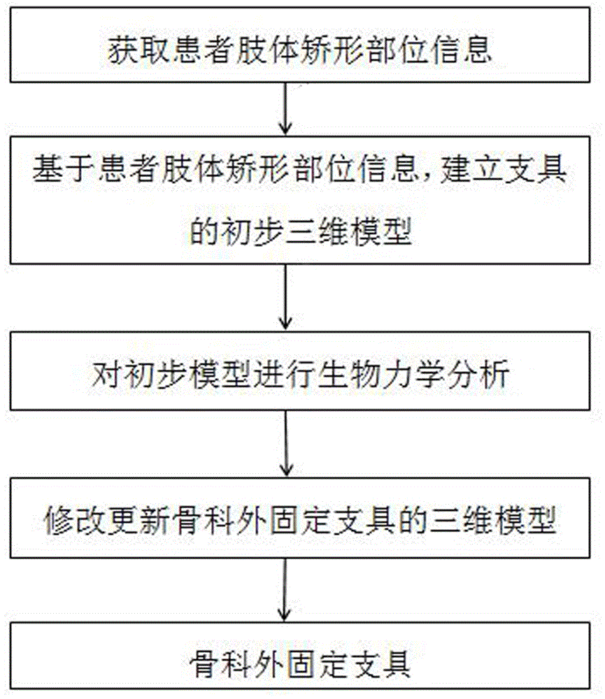
[0026] This embodiment provides a 3D printing preparation method for an orthopedic external fixation brace in order to meet the requirement that the material required for the supporting part of the orthopedic external fixation brace is relatively hard, while the material required for the contact part with soft tissue is relatively soft. Such as figure 1 As shown, the steps are:

[0027] 1) Obtain information on the orthopedic parts of the patient's limbs;

[0028] Since the orthopedic part of the patient's limb is composed of bones and soft tissues, and has a complex structure, it is necessary to use a 3D scanner to accurately obtain the 3D shape information of the orthopedic part of the limb, such as a professional 3D laser scanner or a hand-held 3D scanner. The scanning accuracy of the instrument is better than 0.1mm, and it can accurately obtain the information of the orthopedic parts of the patient's limbs in a short period of time, which is convenient and accurate;

[0...

Embodiment 1

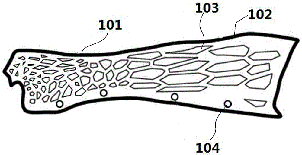
[0044] Such as figure 2 As shown, it is a structural diagram of a wrist fracture fixation brace, which includes a wrist 101 with a relatively high local hardness, a forearm 102 with a relatively low hardness, and a buckle 104 is provided at the opening of the brace; The ratio is small and the hardness is high; the proportion of the hollow 103 at the forearm 102 is large and the hardness is low.

[0045] According to biomechanical analysis, the required Rockwell hardness of the wrist 101 is 1.8, and the Rockwell hardness of the forearm 102 is 1.4 (the Rockwell hardness of the skin is 1.5). The material used in this embodiment is polyethylene, and the softness and hardness are changed by adjusting the filling structure and material filling density of different parts of the physical structure. The area ratio is 40-80%.

[0046] The material used in the brace is added with 2% of the total mass of bamboo charcoal fiber powder and 2% of the total mass of seagull stone, which can ...

Embodiment 2

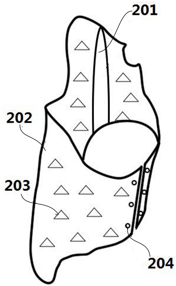
[0049] Such as image 3 As shown, it is a structural diagram of a scoliosis orthopedic brace, including a spine part 201 with relatively high local hardness, a peripheral part 202 with low hardness, and a buckle 204 at the opening of the brace; the spine part 201 is not hollowed out or only There is a small part of hollows; the surrounding part 202 is covered with hollows 203, and the hardness is low. The hardness is changed by changing the ratio of soft and hard materials or the filling rate of the honeycomb structure, and the hardness is higher than that of the peripheral part 202 .

[0050] According to biomechanical analysis, the Rockwell hardness of the required spine part 201 is 2.5, and the Rockwell hardness of the peripheral part 202 is 1.6 (the Rockwell hardness of the skin is 1.5). In this embodiment, the spine part 201 is made of polypropylene, the peripheral part 202 is made of polyvinyl chloride, the area ratio of the hollowed out spine part 201 is 0-20%, and the...


PUM


Abstract

The invention discloses a 3D (three-dimensional) printing preparation method of an orthopaedic external fixing support, which comprises the following steps of: 1, acquiring limb orthopaedic position information of a patient; 2, based on the orthopaedic position information, establishing a primary three-dimensional model of the orthopaedic external fixing support; 3, establishing a physical constitutive model of the orthopaedic external fixing support, and carrying out biomechanical analysis by adopting a finite element method; 4, based on biomechanical analysis, updating primary three-dimensional model data of the orthopaedic external fixing support; 5, based on the updated three-dimensional model data of the orthopaedic external fixing support, designing a final three-dimensional model of the orthopaedic external fixing support, inputting data of the final three-dimensional model into a 3D printing system, and integrally printing and forming to obtain the orthopaedic external fixing support. The 3D printing preparation method disclosed by the invention solves the technical problems of low coincidence degree with a limb orthopaedic position, easiness for causing soft tissue injuries, insufficient antibacterial property, long customizing time and high cost of an external fixing support in the prior art.

Description

technical field [0001] The invention belongs to the technical field of medical devices, and in particular relates to a 3D printing preparation method of an orthopedic external fixation brace. Background technique [0002] The research on orthopedic external fixation has always been the most basic and active field in the treatment of orthopedic patients. From the initial small splint to the invention and application of plaster bandage, thermoplastic splint, detachable foam splint, polymer splint, etc. The treatment of trauma orthopedic patients has led to more effective methods. However, due to the limitations of materials, processes, and costs, the existing orthopedic external fixation braces still have the following problems: (1) The hardness of the brace material in the contact part with the patient's limbs is basically the same, which may be too hard and cause soft tissue damage; (2) The antibacterial property is not enough, and bacteria will grow after wearing for a lon...

Claims


Application Information

Patent Timeline
IPC IPC(8): A61F5/05B33Y10/00B33Y70/00B29C67/00
CPCA61F5/05B33Y10/00B33Y70/00
Inventor 王红滕安军阚生东
Owner QINGDAO UNIQUE PRODS DEV
Why Eureka
Try Eureka