Method for detecting miRNAs based on liquid chip of HCR

A DNA molecule and solution technology, applied in biochemical equipment and methods, microbial determination/inspection, etc., can solve the problems of lack of signal amplification system, inability to complete detection, etc., and achieve the effect of improving sensitivity, high sensitivity and good recovery rate

Active Publication Date: 2016-12-14
SHENZHEN PEOPLES HOSPITAL
View PDF2 Cites 12 Cited by
  • Summary
  • Abstract
  • Description
  • Claims
  • Application Information

AI Technical Summary

Problems solved by technology

It can be seen that due to the omission of the amplification step and the lack of an effective signal amplification...

Method used


Image

  • Method for detecting miRNAs based on liquid chip of HCR
  • Method for detecting miRNAs based on liquid chip of HCR
  • Method for detecting miRNAs based on liquid chip of HCR

Examples


Embodiment 1

[0102] Example 1. Establishment of HCR-based miRNAs liquid-phase chip detection method

[0103] 1. Detection principle of miRNAs liquid chip based on HCR

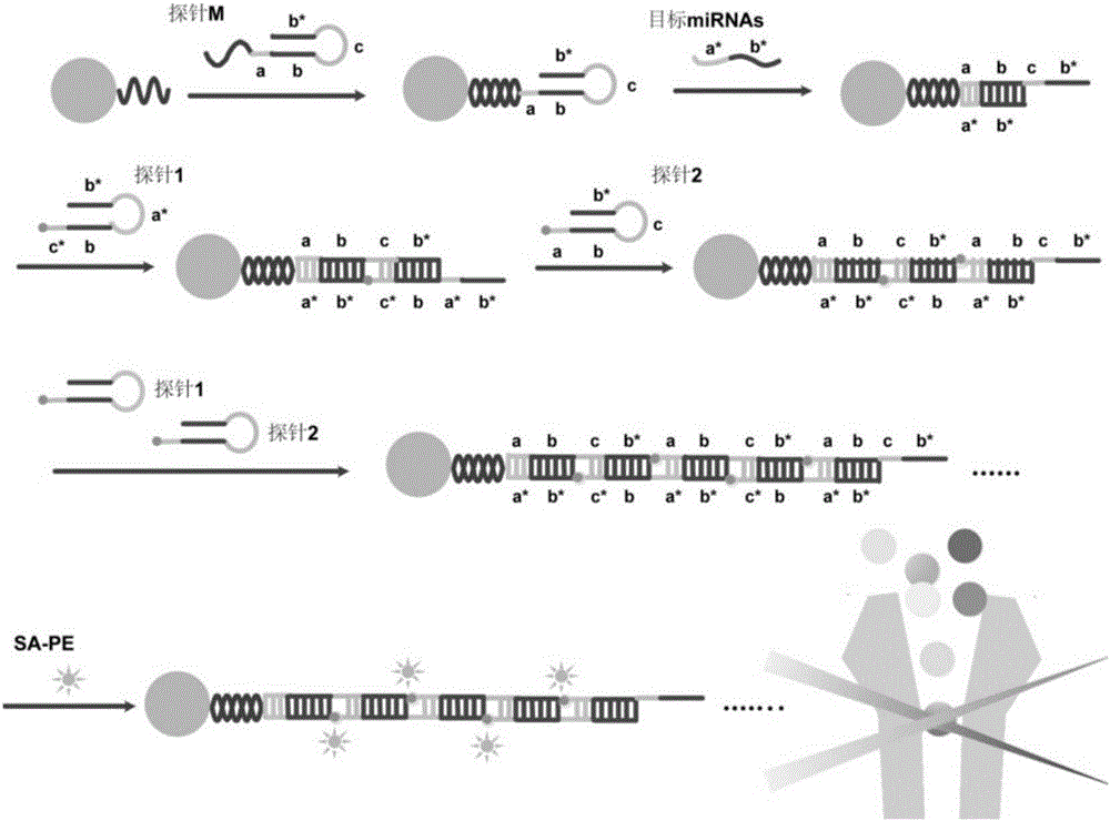
[0104] Schematic diagram of the principle of HCR-based miRNAs liquid chip detection figure 1 .

[0105] Probe M consists of 68 bases, including a sequence that is complementary to the capture sequence on the microsphere; a sequence that is complementary to the target miRNA (a-b); and a sequence that can trigger HCR to initiate a chain reaction (c-b*). After the probe M is denatured and annealed, a stem-loop secondary structure is formed, which is first detected in the system. -TAG TM Microsphere capture. When the target miRNA (a*-b*) is added, the miRNA hybridizes to the probe M, and the hairpin structure of the probe M is opened by a strand displacement reaction, exposing the HCR-triggered chain reaction (c-b* ).

[0106] Both probes 1 and 2 are composed of 44 bases, and are labeled with biotin at the 5' and 3' ends...

Embodiment 2

[0138] Example 2, HCR-based miRNAs liquid chip detection whether it contains let-7a

[0139] Use the miRBase database to query let-7a and miR-21 sequences:

[0140] let-7a: 5'-UGAGGUAGUAGGUUGUAUAGUU-3';

[0141] miR-21: 5'-UAGCUUAUCAGACUGAUGUUGA-3'.

[0142] Let-7a and miR-21 were artificially synthesized, diluted to 100 μM with TE buffer, and stored at -20°C to obtain let-7a standard solution and miRNA21 standard solution.

[0143] 1. Detection of let-7a content by HCR-based miRNAs liquid chip

[0144] 1. Selection of microspheres and design and synthesis of DNA hairpin probes

[0145] 1) Selection of microspheres

[0146] Buy size 12 and size 26 -TAG TM The microspheres were used to detect the content of let-7a and miR-21 respectively. Among them, No. 12 -TAG TM The cross-linked capture sequence on the microspheres is: 5'-AGTAGAAAGTTGAAATTGATTATG-3' (No. 12 microspheres are used to detect let-7a), No. 26 -TAG TM The capture sequence cross-linked by the microsph...

Embodiment 3

[0246] Embodiment 3, detecting target miRNA content in RNA to be tested

[0247] (1) Establish a standard curve

[0248] Dilute 100 μM let-7a standard solution to be tested with buffer A into 6 continuous concentration gradients of 0.1pM, 1pM, 10pM, 100pM, 1nM, 10nM as let-7a standard solution to be tested with different concentrations.

[0249] 1. Selection of microspheres and design and synthesis of DNA hairpin probes

[0250] 1) the selection of microspheres: the same as that of Example 2;

[0251] 2), design and synthesize corresponding probe M, probe 1 and probe 2 according to let-7a and miR-21: the same as that of Example 2;

[0252] 3) Probe pretreatment: the same as that of Example 2;

[0253] 2,

[0254] 1) The probe M is attached to the microsphere to obtain a microsphere-probe complex: the same as that of Example 2;

[0255] 2) The sample to be tested is hybridized with the microsphere-probe complex: the RNA solutions to be tested are respectively 0.1pM, 1pM, 1...


PUM


Abstract

The invention discloses a method for detecting miRNAs based on a liquid chip of HCR and particularly relates to the method for detecting the target miRNAa content in to-be-detected RNA. The method comprises the following steps: (A): (1) respectively designing and synthesizing a probe M, a probe 1 and a probe 2 corresponding to one or more target miRNAs; (2) carrying out HCR reaction on the probe M corresponding to the target miRNA, microspheres corresponding to the target miRNA, the probe 1 and the probe 2, so as to obtain an HCR reactant; and (3) carrying out fluorescent mark detection, determining whether the HCR reactant contains one or more target miRNAs according to the existence of a fluorescence signal in the HCR reactant. The method has the beneficial effects that the sensitivity of a traditional liquid chip is greatly improved, and meanwhile, the aspects of the quantitative range, the specificity and the accuracy degree well presented, so it can be foreseen that a detection measure is provided for the establishment of a relatively rapid, sensitive and simple miRNAs liquid chip detection system for the early-stage new tumor markers.

Description

technical field [0001] The invention relates to the field of biotechnology, in particular to an HCR-based liquid phase chip detection method for miRNAs. Background technique [0002] Although circulating miRNAs are non-invasive tumor markers that can be transformed into clinical practice, many circulating miRNAs with diagnostic value may be overlooked because they are not detected due to the limited sensitivity of most current detection methods, so isolation and detection are useless. miRNAs in cellular body fluid samples remain challenging. In addition, although technologies such as PCR have made significant progress in the detection of single markers, monitoring a single marker is usually not enough for clinical disease diagnosis. Whether it is for early screening of diseases such as cancer or to elucidate the expression patterns of miRNAs in biological systems, it is necessary to be able to detect multiple short-chain miRNAs at extremely low expression levels. But at th...

Claims


Application Information

Patent Timeline
IPC IPC(8): C12Q1/68
CPCC12Q1/6813C12Q1/6851C12Q2525/207C12Q2545/113C12Q2563/107C12Q2525/301C12Q2537/143
Inventor 李富荣杨璐
Owner SHENZHEN PEOPLES HOSPITAL
Why Eureka
Try Eureka