Method for promoting chondrocyte adhesion and tissue engineering cartilage established through method

A chondrocyte and tissue engineering technology, applied in tissue regeneration, medical science, prosthesis, etc., can solve problems such as reducing the survival and proliferation of chondrocytes, affecting the function of the BA technology system, and slowing down the endocytosis of chondrocytes.

Inactive Publication Date: 2017-01-04
ZHONGSHAN HOSPITAL FUDAN UNIV
View PDF2 Cites 2 Cited by
  • Summary
  • Abstract
  • Description
  • Claims
  • Application Information

AI Technical Summary

Problems solved by technology

However, as the culture time prolongs, the endocytosis of biotin by chondrocytes leads to the gradual disappearance of biotin on the surface, which affects the long-term function of the BA technology system.
Lowering the culture temperature to 4°C can slow down the endocytosis of biotin by chondrocytes, but significantly reduce the survival and proliferation of chondrocytes

Method used


Image

  • Method for promoting chondrocyte adhesion and tissue engineering cartilage established through method
  • Method for promoting chondrocyte adhesion and tissue engineering cartilage established through method

Examples


Embodiment 1

[0024] Example 1 Isolation and culture of porcine chondrocytes

[0025] Minipig knee joints were isolated under aseptic conditions, and soft tissues such as muscles, tendons and ligaments around the knee were removed. Irrigate the cartilage surface until no synovial fluid is evident. The surgical blade cut off the cartilage of the medial and lateral femoral condyles in thin slices, paying attention to collecting only the cartilage layer, and stopped cutting when there was bleeding on the surface. Put the cartilage slices into a petri dish, immerse in DMEM and rinse several times, cut into pieces to a size of 1mm×1mm, rinse again in DMEM, remove the supernatant, and transfer the cartilage tissue into a 250mL container containing 0.2% Collagenase Type II (Collagenase Type II, The high-sugar DMEM solution from Sigma Company, USA) was transferred into a constant temperature shaking box (Sanyo, Japan) at 37°C for 4-6 hours, and the cartilage digestion was observed every 2 hours. I...

Embodiment 2

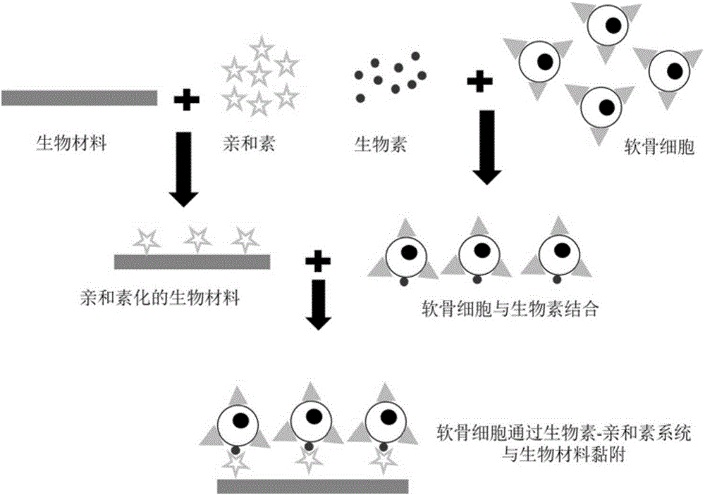
[0026] Example 2 Chitosan Scaffold Avidinization

[0027] The chitosan porous three-dimensional scaffold is prefabricated into a cylinder with a diameter of 10 mm and a height of 3 mm, the porosity is >90%, and the pore diameter is 100-150 μm, and it is sterilized by ethylene oxide for use. Take the above-mentioned chitosan scaffold and place it in a 12-well culture plate, add 3.0 mL of 0.1 mg / mL avidin solution, react on a shaker (125 r / min) at room temperature for 1 h, and use 200 mg / mL double antibody and 25 mg / mL amphotericin Primer B was washed twice with PBS and dried naturally for later use.

Embodiment 3

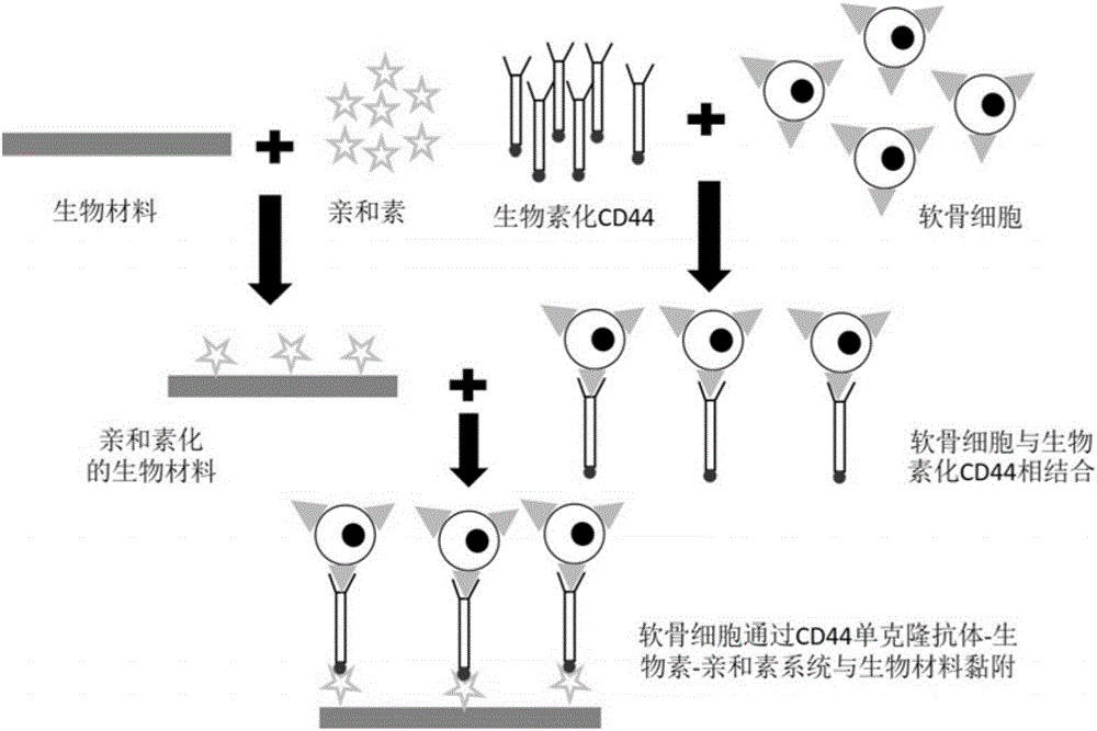
[0028] Example 3 Construction of tissue engineered cartilage in vitro by CD44 monoclonal antibody-biotin-avidin system

[0029] Chondrocytes at 5×10 7 cells / mL density and biotinylated monoclonal antibody CD44 after static reaction for 30min (0.25μg biotinylated monoclonal antibody CD44 / 1×10 6 cells) were washed twice with PBS, and then inoculated in a 24-well plate with chitosan scaffolds, inoculated with 200 μL cell suspension in each scaffold, and placed at 37 ° C, 5% CO 2 Culture in an incubator to further combine cells and scaffolds. 4 hours after inoculation, transfer the cell-scaffold complex to a new 24-well plate, gently add 2 mL of culture medium and let it stand in an incubator for culture, and change the medium every other day.


PUM

PropertyMeasurementUnit
pore sizeaaaaaaaaaa
diameteraaaaaaaaaa
heightaaaaaaaaaa

Abstract

The invention discloses a method for promoting chondrocyte adhesion and tissue engineering cartilage established through the method. According to the method, a monoclonal antibody of a chondrocyte surface high-expression antigen is coupled with a biotin-avidin system, and on the one hand, the problem of endocytosis of chondrocyte in the biotin-avidin system to biotins is avoided; on the other hand, by means of high binding force of the antigen antibody, the adhesion capacity of the chondrocyte to a bio-material is improved, proliferation of tissue engineering cartilage seed cells and expression of chondrocyte types are promoted, then the tissue engineering cartilage is established, and the effect of repairing cartilage defects is improved.

Description

technical field [0001] The invention relates to a method for promoting chondrocyte adhesion and tissue engineering cartilage constructed therefrom. Background technique [0002] The repair of articular cartilage defects is a difficult point in clinical orthopedics. The rise of tissue engineering techniques has shed new light on addressing this issue. But there is still a big gap between its repair effect and normal cartilage. According to the analysis, the adhesion between the seed cells and the scaffold carrier is not strong, the seed cells fall off from the scaffold, and the insufficient number of seed cells may be one of the main reasons leading to this unsatisfactory effect. In living tissues, the adhesion of cells and extracellular matrix is ​​accomplished through the combination of receptors on the cell membrane and adhesion proteins on the extracellular matrix. Apparently, tissue engineering scaffolds lack such adhesion proteins. Therefore, cells can only attach t...

Claims


Application Information

Patent Timeline
Patent Type & Authority Applications(China)
IPC IPC(8): A61L27/38A61L27/50
CPCA61L27/3817A61L27/3852A61L27/3895A61L27/50A61L2430/06
Inventor 林红周健陈峥嵘王会仁曹露马易群郭常安董健
Owner ZHONGSHAN HOSPITAL FUDAN UNIV
Why Eureka
Try Eureka