Efficient CIK cell preparation and detection method

A cell and efficient technology, applied in biochemical equipment and methods, animal cells, vertebrate cells, etc., can solve the problems of low CD3+CD56+ double positive expression rate, unstable preparation process, and low proliferation multiple of CIK cells

Inactive Publication Date: 2017-01-04
中卫华医(北京)生物科技有限公司 +1
View PDF4 Cites 10 Cited by
  • Summary
  • Abstract
  • Description
  • Claims
  • Application Information

AI Technical Summary

Problems solved by technology

The purpose of the present invention is to improve the traditional CIK cell preparation method in view of the problems of low proliferat...

Method used


Image

  • Efficient CIK cell preparation and detection method
  • Efficient CIK cell preparation and detection method
  • Efficient CIK cell preparation and detection method

Examples


preparation example Construction

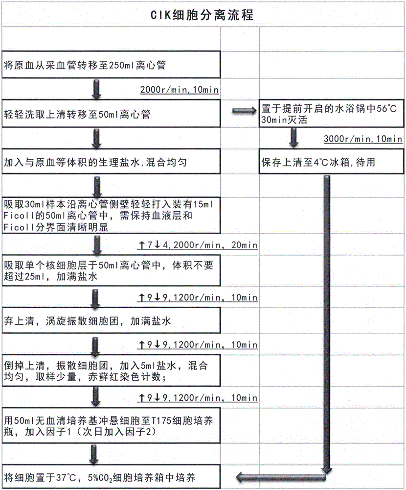
[0099] The method for preparing high-efficiency CIK cells of the present invention comprises the following steps:

[0100] (1) Whole blood test and blood sample storage: Use a pipette to transfer the original blood from the blood collection tube to a centrifuge tube, blow and beat repeatedly to mix well: take out the blood sample for microbial detection, and store the blood sample in a cryopreservation tube for refrigerated storage. As a file for subsequent inspection;

[0101] (2) Plasma treatment: Put the centrifuge tube containing the original blood into the centrifuge, balance and centrifuge, transfer the upper layer plasma to the centrifuge tube, close the lid tightly, stick the sealing film, and place it in a 56°C water bath to inactivate , after inactivation, the pellet was discarded by centrifuge, and the supernatant FicoLL was saved for later use;

[0102] (3) Separation of mononuclear cells: add an equal volume of normal saline to the original blood, pipette to dilu...

Embodiment 2

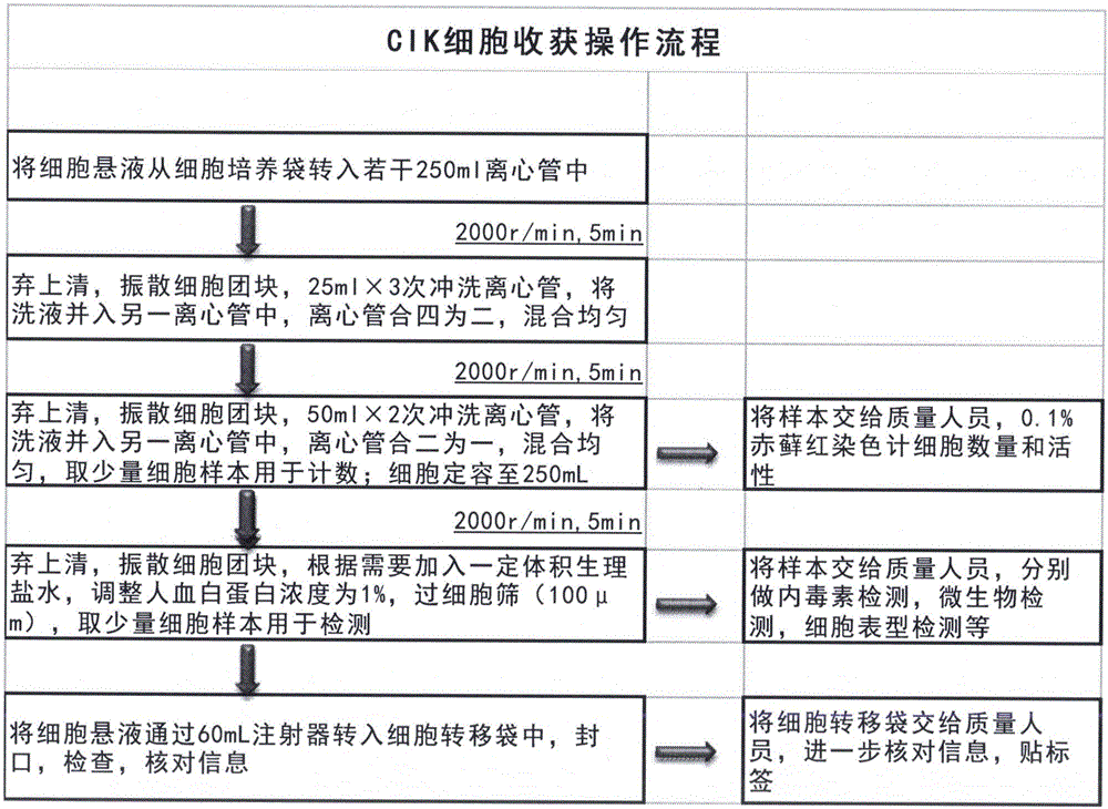
[0203] Embodiment 2, the difference between the high-efficiency CIK cell preparation and detection method of the present invention and the above-mentioned embodiment 1 is only: 4.1 In the detection of cell number and activity: the number of cells requires 3-4×10 9; 4.2 Cell sterility test: observe the result after 48 hours, if the culture medium is still clear, the result is negative. 4.5 Cell phenotype detection: Wash once with normal saline.

[0204] Embodiment 2, the method for preparing and detecting high-efficiency CIK cells of the present invention differs from the above-mentioned embodiment 1 only in that: 4.1 In the detection of cell number and activity: the number of cells requires 5-6×10 9 ; 4.2 Cell sterility test: observe the result after 72 hours, if the culture medium is still clear, it means the result is negative. Wash with normal saline twice.

[0205] For the above examples, please refer to the attached CIK cell preparation flow chart.

[0206] The present...


PUM


Abstract

The invention relates to an efficient CIK cell preparation and detection method and aims at solving the problems of few proliferation times, unstable preparation process and the like of the prior art. The efficient CIK cell preparation and detection method comprises the following steps that sterile acquisition is conducted peripheral blood of a healthy person or a patient according to a lymphocyte absolute value; whole blood testing and blood sample preservation are performed; separation and inactivation of blood plasma are performed; a density gradient centrifugation method is adopted, namely a single mononuclear cell is separated out by using lymphocyte separation liquid Ficoll; in the CIK cell induction process, a factor 1 and a factor 2 are added successively, collection can be performed only through serum-free induced cell multiplication culture for about 2-3 weeks, and a CIK cell preparation is prepared. The method has the advantages of being small in blood collecting quantity, stable in preparation process, simple and convenient to operate and high in cell proliferation times and cytotoxic activity.

Description

technical field [0001] The invention relates to a method for preparing CIK cells, in particular to a method for preparing and detecting highly efficient CIK cells. [0002] Background of the invention [0003] Adoptive immunotherapy has become the fourth treatment method after the three traditional treatment methods such as surgery, radiotherapy and chemotherapy, especially Cytokine-Induced Killer cells (CIK), because of its rapid proliferation , wide antitumor spectrum, high antitumor activity, low toxicity to normal bone marrow hematopoietic precursor cells, sensitive to multi-drug resistant tumor cells, and can be combined with traditional treatment methods. Cell activity is strong, an ideal effector cell suitable for clinical application. [0004] CIK cells are extremely rare in normal peripheral blood, only 1% to 5%. The discovery of CIK cell preparation technology makes it possible to apply it clinically. CIK cells were first reported by American scientists in the 19...

Claims


Application Information

Patent Timeline
IPC IPC(8): C12N5/0783C12Q1/02G01N33/579G01N33/68
CPCC12N5/0646C12N5/0636C12N2501/2301C12N2501/2302C12N2501/24C12N2501/51C12N2501/515G01N33/48735G01N33/5005G01N33/6854
Inventor 谷广其王晶李亚平邱丽媛赵进军何文丽
Owner 中卫华医(北京)生物科技有限公司
Why Eureka
Try Eureka