
Preparation method of nano Gd-MOFs for magnetic resonance imaging
A kind of magnetic resonance imaging and nanotechnology, applied in the direction of nanotechnology for sensing, preparations for in vivo experiments, nanotechnology, etc., can solve the adverse effects of MRI contrast agent use, Gd-MOFs nanorod toxicity, application Obstacles and other problems, to achieve obvious effect, large relaxation rate, good biocompatibility
- Summary
- Abstract
- Description
- Claims
- Application Information
AI Technical Summary
Problems solved by technology
Method used
Image

Examples
Embodiment Construction
[0033] Embodiments of the present invention will be described in detail below in conjunction with examples. Those skilled in the art will understand that the following examples are only preferred examples of the present invention, so as to better understand the present invention, and thus should not be considered as limiting the scope of the present invention.
[0034] The experimental methods in the following examples, unless otherwise specified, are conventional methods; the experimental materials used, unless otherwise specified, were purchased from conventional biochemical reagent manufacturers.
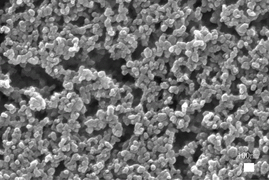
[0035] In the following examples, a desktop high-speed centrifuge (XiangYi TG16-WS) was used for centrifugation; the SEM scanning electron microscope photos were obtained by using a Quanta FEG 250 field emission environment scanning electron microscope.
[0036] (1) Dissolve 0.1mmol (20mg) trimellitic acid in 8ml DMF, fully dissolve, and record it as solution A; mix 1mmol 1-ethyl...
PUM
 Abstract
Description
Claims
Application Information