Cell separating, preparation and dyeing integrated device and circulating tumor cell capturing method

A tumor cell and cell technology, applied in the field of in vitro diagnosis and microfluidics, can solve the problems of low detection efficiency, expensive reagents, and large limitations of antibody capture cells, achieve less sample consumption, avoid false positives and false negatives, Ease of integration and miniaturization

Pending Publication Date: 2017-07-28
SUZHOU WENHAO MICROFLUIDIC TECH CO LTD
View PDF10 Cites 4 Cited by
  • Summary
  • Abstract
  • Description
  • Claims
  • Application Information

AI Technical Summary

Problems solved by technology

Although the counting has high sensitivity, there are expensive instruments and reagents consumed, and the limitation

Method used


Image

  • Cell separating, preparation and dyeing integrated device and circulating tumor cell capturing method
  • Cell separating, preparation and dyeing integrated device and circulating tumor cell capturing method
  • Cell separating, preparation and dyeing integrated device and circulating tumor cell capturing method

Examples


Embodiment Construction

[0033] Circulating Tumor Cells: Tumor cells that have spread from a primary tumor into the peripheral blood circulation.

[0034] The technical solutions in the embodiments of the present invention will be described in detail below in conjunction with the accompanying drawings in the embodiments of the present invention. Obviously, the described embodiments are only some of the embodiments of the present invention, not all of them. Based on the embodiments of the present invention, all other embodiments obtained by persons of ordinary skill in the art without making creative efforts belong to the protection scope of the present invention.

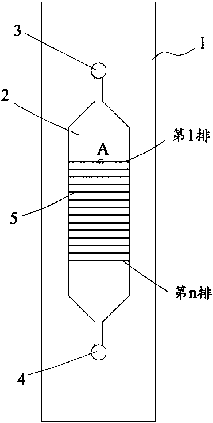
[0035] combine figure 1 and figure 2 As shown, the integrated device for cell separation, preparation and staining includes a substrate 1 and a channel 2 extending in the substrate along a first direction, and a sample inlet 3 and a sample outlet 4 are respectively formed at both ends of the communication 2 located in the first direction,...


PUM


Abstract

The invention discloses a cell separating, preparation and dyeing integrated device and a circulating tumor cell capturing method. A chip comprises a base material and a channel which extends into the base material, wherein a sample inlet and a sample outlet are respectively formed in two ends of the channel; n rows of micro-column arrays are arranged in the channel in an arrayed manner, and n is greater than or equal to 2; each row of micro-column arrays respectively comprises a plurality of micro-columns which are arranged at intervals; a capturing gap is formed between each two adjacent micro-columns, and the following conditions are met: (a), the capturing gaps of an xth row of micro-column arrays are greater than the capturing gaps of a (x+1)th row of micro-column arrays from the sample inlet to the sample outlet, and x is greater than or equal to 1 and smaller than n; and (b), the capturing gaps of the nth row of micro-column arrays are smaller than the outer diameters of circulating tumor cells. The method has the advantages of small amount of consumed samples, high resolution precision and sensitivity, easiness in integration and miniaturization and the like. In addition, the circulating tumor cells are captured on the basis of the characteristic that the boundary dimension of the circulating tumor cells is different from that of blood corpuscles, and capturing of the circulating tumor cells does not depend on expression of an EpCAM antibody.

Description

technical field [0001] The application belongs to the field of in vitro diagnosis and microfluidic technology, and in particular relates to an integrated device for cell separation, preparation and staining and a method for capturing circulating tumor cells. Background technique [0002] Circulating tumor cells (CTCs) refer to tumor cells that have diffused from the primary tumor into the peripheral blood circulation system, and their appearance indicates that tumor cells have begun to spread from the lesion to the surroundings, which may lead to more organs and tissues. produce lesions. [0003] Circulating tumor cells can appear in the early stage of tumorigenesis, and studies have shown that about 90% of cancer death cases are related to the spread of CTCs. Therefore, CTCs detection is important in the early prevention of major diseases, evaluation of treatment effects, drug sensitivity testing, and tumor recurrence monitoring. It is of great significance in medical appl...

Claims


Application Information

Patent Timeline
IPC IPC(8): G01N1/28G01N1/34G01N1/31
CPCG01N1/28G01N1/31G01N1/34
Inventor 冯澄宇
Owner SUZHOU WENHAO MICROFLUIDIC TECH CO LTD
Why Eureka
Try Eureka