Automatic-focusing microscopic endoscopic fluorescence imaging system

A technology of automatic focusing and fluorescence imaging, which is applied in the direction of endoscopy, using fluorescence emission for analysis, using spectrum diagnosis, etc., can solve the problems of time-consuming and laborious, and achieve the effect of convenient equipment maintenance, convenient use, and simple realization

Inactive Publication Date: 2017-08-18
苏州双威医疗器械科技有限公司
View PDF8 Cites 11 Cited by
  • Summary
  • Abstract
  • Description
  • Claims
  • Application Information

AI Technical Summary

Problems solved by technology

[0011] High-resolution fluorescence imaging (HRME) uses manual focusing, which is time-consuming and laborious

Method used


Image

  • Automatic-focusing microscopic endoscopic fluorescence imaging system
  • Automatic-focusing microscopic endoscopic fluorescence imaging system

Examples


Embodiment

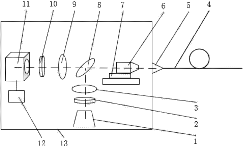
[0047] Taking the fluorescence microscopic imaging of rabbit digestive tract mucosa as an example, the working method of the present invention is introduced

[0048] 1. According to the spectral characteristics of the fluorescent contrast agent acriflavine hydrochloride used, select the appropriate excitation light filter (455nm) and emission light filter (525nm), dichroic mirror filter (505nm) combination.

[0049] 2. Install the imaging fiber on the fiber coupler, turn on the light source and the camera, and stick the front end of the imaging fiber to the gauze stained with acridine yellow hydrochloride. The white bright spot displayed on the screen means that the proximal end of the imaging fiber is not zooming in. on the focal plane of the objective lens.

[0050] 3. An image data processing control module is set, and the focus position is searched by evaluating the quality of the collected images by using the auto-focus evaluation function. The motor drives the platform...


PUM


Abstract

The invention provides an automatic-focusing microscopic endoscopic fluorescence imaging system. The system comprises a white-light LED light source, a scientific-grade low-temperature refrigeration CCD camera or an industrial-grade CCD camera, a small-diameter flexible image transmitting optical fiber and an optical fiber outer sleeve, an amplifying objective lens and an automatic focusing device, an exciting light filter, an emission light filter, a dichroscope light filter, an optical fiber collimation coupler, a micromotion translation table, an image data processing control module and a device shell. According to the system, in combination with the imaging principle and the automatic focusing technology of the fluorescence microscope, by optimizing hardware equipment, developing image processing software and constructing the automatic-focusing microscopic endoscopic imaging device, the device as the sub-mirror is fused with the existing digestive endoscopy, and then the brand-new bimodal endoscopic imaging mode realizing fluorescence microscopic molecular imaging and special light anatomic construction imaging is established.

Description

technical field [0001] The invention relates to imaging technology, in particular to an automatic focusing microendoscopic fluorescence imaging system. Background technique [0002] Digestive system tumors are among the most common tumors worldwide. Early detection, early treatment, and improving the diagnosis level of early cancer have far-reaching significance for improving the survival rate of patients and reducing social and economic burdens. Current international research shows that endoscopy is the most effective way to find digestive system tumors. However, existing endoscopic techniques have many problems, such as low detection rate and high missed diagnosis rate. To solve existing problems, molecular imaging provides us with new ideas. Molecular imaging studies biological processes in vivo at the cellular and molecular levels, and at the same time uses targeted probes combined with specific molecules to achieve real-time, quantitative imaging. Digestive endoscop...

Claims


Application Information

Patent Timeline
Patent Type & Authority Applications(China)
IPC IPC(8): A61B1/04A61B1/06A61B5/00A61B1/00
CPCA61B1/00009A61B1/00131A61B1/00165A61B1/043A61B1/0684A61B5/0071A61B5/0075A61B5/0084A61B2503/40
Inventor 屈亚威付甲龙刘海峰
Owner 苏州双威医疗器械科技有限公司
Why Eureka
Try Eureka