

A kind of staining agent for rapid color development after cell staining in urine and its application method
A dye and cell technology, applied in the field of in vitro diagnosis, can solve the problems of complex preparation process, long dyeing time, easy aggregation, etc., and achieve the effects of simple operation, high detection efficiency, and short color development time.
- Summary
- Abstract
- Description
- Claims
- Application Information
AI Technical Summary
Problems solved by technology
Method used
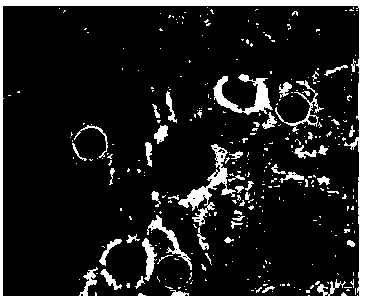
Image


Examples
Embodiment 1
[0064] 1. Preparation of staining agent Reagent A:
[0065] Add 5.00g of methanol, 95.00g of ethylene glycol and 1.20g of neutral red to the brown reagent bottle in order to obtain the first mixed solution A1; after that, place the brown reagent bottle containing the above-mentioned first mixed solution A1 on the shaker , Dissolved at a medium speed for 3 hours, so that the drug was completely dissolved, and the second mixed solution A2 was obtained; then, the above-mentioned second mixed solution A2 was filtered with a 0.22um filter membrane, and the filtered solution was marked as reagent A, and placed in a brown bottle save in .
[0066] Reagent B:
[0067] Add 100.00g of purified water, 4.20g (200mM) of 1,3-bis[(tris(hydroxymethyl)methylamino]propane (Bis-tris), and 8.00g of sodium propionate into the reagent bottle, and fully dissolve to obtain the first A mixed solution B1; add propionic acid to the first mixed solution B1 to make the pH of the solution B1 within the r...
Embodiment 2
[0071] 1. Preparation of staining agent Reagent A:
[0072] Add 100.00g of ethylene glycol and 1.20g of neutral red to the brown reagent bottle in sequence to obtain the first mixed solution A1; after that, place the brown reagent bottle containing the above first mixed solution A1 on a shaker and dissolve at a medium speed After 3 hours, the drug was completely dissolved to obtain the second mixed solution A2; then, the above-mentioned second mixed solution A2 was filtered with a 0.22um filter membrane, and the filtered solution was marked as reagent A, and stored in a brown bottle.
[0073] Reagent B:
[0074] Add 100.00g of purified water, 2.10g (50mM) of 2-(N-morpholine)ethanesulfonic acid monohydrate (MES), 4.00g of sodium propionate, 4.00g of EDTA-K into the reagent bottle 2 After fully dissolving, the first mixed solution B1 is obtained; the pH value of the first mixed solution B1 is adjusted to within the range of 6.00±0.05 (25±1°C) with propionic acid; Filter, label...
Embodiment 3
[0078] 1. Preparation of staining agent Reagent A:
[0079] Add 100g of glycerol and 0.6g of neutral red to the brown reagent bottle in sequence to obtain the first mixed solution A1; after that, place the brown reagent bottle containing the above-mentioned first mixed solution A1 on a shaker and dissolve it at a medium speed for 3.5 hours, the medicine was completely dissolved to obtain the second mixed solution A2; then the above-mentioned second mixed solution A2 was filtered with a 0.22um filter membrane, and the filtered solution was marked as reagent A, and placed in a brown bottle for preservation.
[0080] Reagent B:
[0081] Add 100g of purified water, 0.61g (50mM) of Tris (Tris), 8g of sodium propionate and 1.9g of citric acid into the reagent bottle, and fully dissolve to obtain the first mixed solution B1; Adjust the pH value of a mixed solution B1 to within the range of 6.70±0.05 (25±1°C); then use a 0.22 μm filter membrane to filter the above-mentioned second mixe...
PUM
 Abstract
Description
Claims
Application Information