Acute cerebral ischemia image segmentation model acquisition method and acute cerebral ischemia image segmentation method

An acute cerebral ischemia and image segmentation technology, applied in the field of medical image processing, can solve the problems of time-consuming and energy-consuming, difficult to determine the accurate position, and large position changes, so as to reduce the calculation time, increase the accuracy, and improve the accuracy. Effect

Inactive Publication Date: 2017-10-13
杭州全景医学影像诊断有限公司
View PDF4 Cites 11 Cited by
  • Summary
  • Abstract
  • Description
  • Claims
  • Application Information

AI Technical Summary

Problems solved by technology

However, due to the rapid onset of acute cerebral ischemia, large changes in location, and difficulty in determining the shape, it is difficult to have a general segmentation method, and manual segmentation requires medical experience and anatomical knowledge, which is relatively subjective and takes time and effort.
[0005] In recent years, researchers have proposed some segmentation methods for cerebral ischemia, but there are still relatively few studies on acute cerebral ischemia within 6 hours, because compared with other general ischemia, acute cerebral ischemia has less signal in MR images. The difference is relatively large, and the boundary is blurred, so it is difficult to d

Method used


Image

  • Acute cerebral ischemia image segmentation model acquisition method and acute cerebral ischemia image segmentation method
  • Acute cerebral ischemia image segmentation model acquisition method and acute cerebral ischemia image segmentation method
  • Acute cerebral ischemia image segmentation model acquisition method and acute cerebral ischemia image segmentation method

Examples


Embodiment Construction

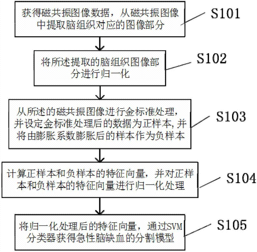
[0047] Please refer to figure 1 As shown, the acute cerebral ischemia image segmentation model of the present invention obtains a process flow chart, specifically including:

[0048] S101: Acquire the magnetic resonance image data and extract the brain tissue, binarize the magnetic resonance image, extract the largest connected region which is the mask image of the brain tissue, multiply the mask image and the magnetic resonance image to obtain the brain tissue image ;

[0049] S102: Normalize the extracted brain tissue images;

[0050] S103: Draw the ischemic region on the extracted brain tissue image as a positive sample, and use the data expanded by the positive sample according to the set expansion coefficient as a negative sample;

[0051] S104: Extract feature vectors from positive samples and negative samples, and normalize feature vectors;

[0052] S105: The normalized feature vector is used to obtain an acute cerebral ischemia segmentation model through an SVM clas...


PUM


Abstract

The invention provides an acute cerebral ischemia image segmentation model training method and an acute cerebral ischemia image segmentation method. The method comprises steps that magnetic resonance image data is acquired, and brain tissue images are extracted; normalization operation of the extracted brain tissue images is carried out; ischemic areas of the extracted brain tissue images are acquired as a positive sample, and expansion of the positive sample is carried out according to a set expansion coefficient to acquire data which is taken as a negative sample; characteristic vectors are respectively extracted from magnetic resonance images T2, DWI and ADC, the positive sample and the negative sample of asymmetric maps ASM, and normalization processing on the characteristic vectors is carried out; based on the characteristic vectors after normalization processing, an acute cerebral ischemia segmentation model is acquired through an SVM classifier. The method is advantaged in that the provided acute cerebral ischemia segmentation model can be applied to ischemic area segmentation, a segmentation speed is fast, and high accuracy is realized.

Description

technical field [0001] The invention relates to the field of medical image processing, in particular to a method for obtaining an acute cerebral ischemia image segmentation model and a method for acute cerebral ischemia image segmentation. Background technique [0002] Cerebrovascular disease is the second leading cause of death in the world. Ischemic cerebrovascular disease accounts for about 80% of all cerebrovascular diseases. According to the third national cause of death survey released by the Ministry of Health in 2008, cerebrovascular diseases have surpassed malignant tumors to become the number one cause of death in China. Acute cerebral ischemia refers to the clinical symptoms of brain dysfunction caused by insufficient blood supply to a certain part of the human brain, leading to corresponding brain function defects. Acute cerebral ischemia has an acute onset and is very harmful. Most patients will experience symptoms that affect their quality of life, such as he...

Claims


Application Information

Patent Timeline
IPC IPC(8): G06T7/11G06K9/62
CPCG06T7/11G06T2207/20081G06T2207/10088G06T2207/20104G06F18/2411
Inventor 彭雨晴李成州许远帆王石峰宫健
Owner 杭州全景医学影像诊断有限公司
Why Eureka
Try Eureka