Method for separating and purifying CD8+T cells from liver cancer tissue

A technology for separating and purifying liver cancer tissues, applied in cell dissociation methods, biochemical equipment and methods, tissue culture, etc., can solve the unavoidable non-specific binding of cell debris and dead cells, low sorting purity, and low cell yield To improve the purity and efficiency of sorting, the cell yield is not affected, and the sorting cell yield is low.

Inactive Publication Date: 2017-11-28
MENGCHAO HEPATOBILIARY HOSPITAL OF FUJIAN MEDICAL UNIV
View PDF3 Cites 5 Cited by
  • Summary
  • Abstract
  • Description
  • Claims
  • Application Information

AI Technical Summary

Problems solved by technology

These two methods have their own advantages and disadvantages in practical application: (1) Although the separation purity of flow separation can reach 90%, the equipment is expensive, the operation is complicated, the separation time is long, and the high-purity mode is selected for separation. There will inevitably be a loss of many cells during the selection, resulting in a final cell yield of less than 10^5
(2) Tumor tissue is complex and contains many different types of cells. At the same time, the process of digesting the tissue

Method used


Image

  • Method for separating and purifying CD8+T cells from liver cancer tissue
  • Method for separating and purifying CD8+T cells from liver cancer tissue
  • Method for separating and purifying CD8+T cells from liver cancer tissue

Examples


Embodiment 1

[0038] 1. Materials, methods and main reagent consumables

[0039] (1) Specimens: The clinical data of Fuzhou Infectious Disease Hospital who will undergo surgical resection of primary hepatocellular carcinoma were analyzed through the hospital's medical record system. After the approval of the hospital ethics committee and the informed consent of the patients themselves or their families, surgically resected tumor tissues were collected. It is planned to select 4 cases, and the inclusion criteria are solitary primary hepatocellular carcinoma patients with a tumor diameter of 5-10 cm; the exclusion criteria are patients with combined cholangiocarcinoma and liver metastases; Cases such as B-ultrasound-guided radiofrequency therapy or other treatments. The preoperative examination items of the enrolled cases included liver B-ultrasound, CT or MRI, blood liver function, blood AFP, lung CT, brain CT, whole body bone ECT, blood hepatitis B two and a half, etc. All enrolled cases ...

Embodiment 2

[0066] The method for separating and purifying CD8+ T cells from human liver cancer tissue by Miltenyi immunomagnetic bead sorting comprises the following steps:

[0067] 1. Separation of tumor infiltrating lymphocytes (with step 1 of Example 1)

[0068] 2. CD8+ T cell MACS sorting (same as step 3 of Example 1)

[0069] 3. Detection of cell purity and cell number

[0070] (1) Take 100ul of cell suspension and add 5ul of CD8 and CD3 antibodies at the same time, incubate at room temperature in the dark for 25min, wash and centrifuge twice, and resuspend the cells in 200ul of PBS;

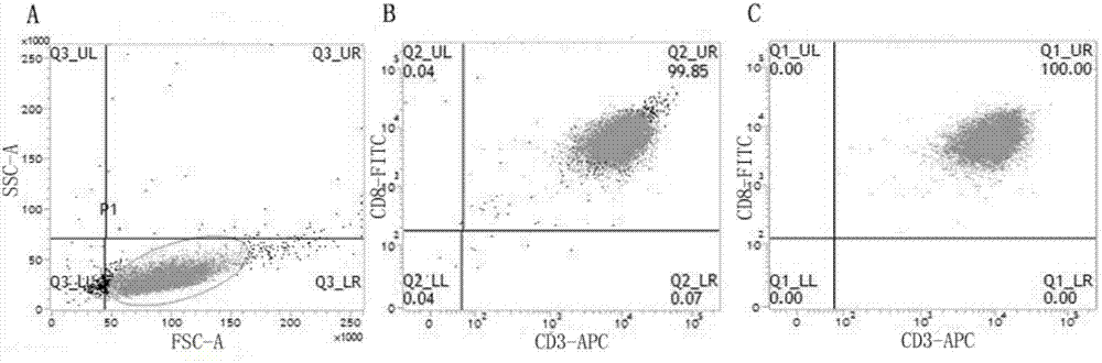
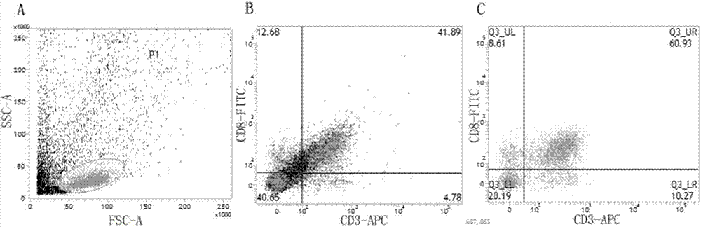
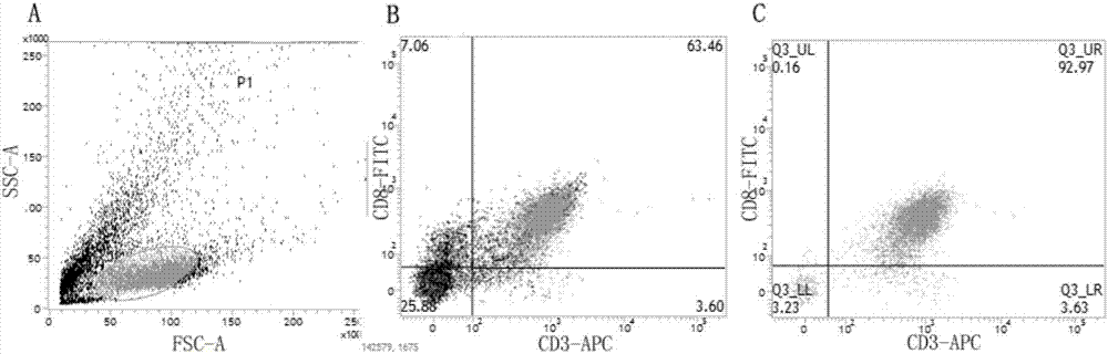
[0071] (2) The cell purity detected by BD flow cytometry is lower than 50% (results see figure 2 );

[0072] (3) Take 20ul of cell suspension, and measure the number of cells with a cell counter, and the number of cells is greater than 3×10 5 .

Embodiment 3

[0074] The method for separating CD8+ T cells from human liver cancer tissue by Ficoll density gradient centrifugation combined with magnetic bead sorting, the specific steps are as follows:

[0075] 1. Separation of tumor infiltrating lymphocytes (with step 1 of Example 1)

[0076] 2. Density Gradient Centrifugation

[0077] (1) Slowly add 4ml of Ficoll solution and 8ml of cell suspension to a 15ml centrifuge tube;

[0078] (2) Adjust centrifuge ascending speed to 1, descending speed to 0, and centrifuge at 800g for 20min;

[0079] (3) Carefully absorb the cells on the upper layer of Ficoll, wash the cells twice with PBS, and collect the cells.

[0080] 3. CD8+ T cell MACS sorting (same as step 3 of Example 1)

[0081] 4. Detection of cell purity and cell number

[0082] (1) Take 100ul of cell suspension and add 5ul of CD8 and CD3 antibodies at the same time, incubate at room temperature in the dark for 25min, wash and centrifuge twice, and resuspend the cells in 200ul of...


PUM


Abstract

The invention relates to a method for separating and purifying CD8+T cells from liver cancer tissue. The method for separating the CD8+T cells comprises the following specific steps: (1) separating tumor infiltration immune cells; (2) performing discontinuous density gradient centrifugation with 30% percoll and 60% percoll; and (3) sorting the CD8+T cells by using MACS (Magnetic Activated Cell Sorting) microbeads. With the combination of density gradient centrifugation and microbead sorting, the CD8+T cells are successfully purified from liver cancer tissue, the purity of the cells is identified by using a flow cytometry, the purity is up to 95% or greater, and meanwhile the cell number is up to 3*10<5> or higher.

Description

(1) Technical field [0001] The invention relates to a method for separating and purifying CD8+ T cells from liver cancer tissue. (2) Background technology [0002] CD8+ T lymphocytes are the main effector cells of anti-tumor immunity. CD8+ T cells can recognize endogenous antigenic peptides presented by MHC class I molecules, and can produce IFN-γ to mediate the killing of target cells carrying antigens. In addition, there are CD8+ T cells that can produce IL-17 in tumor tissue, and the secretion of IL-17 can promote the further development of tumor by promoting blood vessels, and at the same time, tumor-activated monocytes can secrete a series of key cytokines (IL -1β, IL-6 and IL-23) to induce the proliferation of IL-17-secreting CD8+ T cells. It can be seen that CD8 + There is a relatively complex interaction between T cells and the tumor microenvironment, which can promote the immune response to kill tumor cells, or inhibit the immune response and cannot effectively k...

Claims


Application Information

Patent Timeline
IPC IPC(8): C12N5/0783
CPCC12N5/0636C12N2509/00C12N2509/10
Inventor 刘景丰刘小龙董秀清蔡志雄陈耕李振丽
Owner MENGCHAO HEPATOBILIARY HOSPITAL OF FUJIAN MEDICAL UNIV
Why Eureka
Try Eureka