Electromagnetic-acoustic orthopedic surgery guiding device and alarm method thereof

A technology of orthopedic surgery and guiding devices, which is applied in the field of ultrasonic sensors and auxiliary drilling instruments, electromagnetic acoustic orthopedic surgical guiding devices and their alarms, can solve the problems of huge equipment, pedicles cannot be continuously monitored, and expensive, etc., to achieve Overcome the effect of sensor damage by force

Pending Publication Date: 2018-01-09
SHANGHAI JIAO TONG UNIV
View PDF11 Cites 0 Cited by
  • Summary
  • Abstract
  • Description
  • Claims
  • Application Information

AI Technical Summary

Problems solved by technology

The advantages of the first method are simplicity, low cost, and good safety; the disadvantage is that it relies more on the clinical experience of doctors
The advantage of the second method is that it can prevent complications such as nerve root damage caused by improper instrument position, and it is more sensitive than X-ray fluoroscopy; the disadvantage is that it cannot continue to monitor after the pedicle is pierced, and it cannot directly reflect the screw position
The third method includes X-ray image and ultrasonic image monitoring. The advantage is that the position of the nail can be judged intuitively, which can greatly improve the success rate of the operation; Equipment, the operation is complicated, the operation time is relatively long, and if X-images are used, it has radiation
Such as David T.Raphael, Jin Ho Chang, Yao Ping Zhang, David Kudija, Thomas C.Chen, K.Kirk Shung proposed "A-Mode ultra...

Method used


Image

  • Electromagnetic-acoustic orthopedic surgery guiding device and alarm method thereof
  • Electromagnetic-acoustic orthopedic surgery guiding device and alarm method thereof
  • Electromagnetic-acoustic orthopedic surgery guiding device and alarm method thereof

Examples


Embodiment Construction

[0051] The present invention will be described in detail below with reference to the drawings and specific embodiments. This embodiment is implemented on the premise of the technical solution of the present invention, and a detailed implementation mode and specific operation process are given, but the protection scope of the present invention is not limited to the following embodiments.

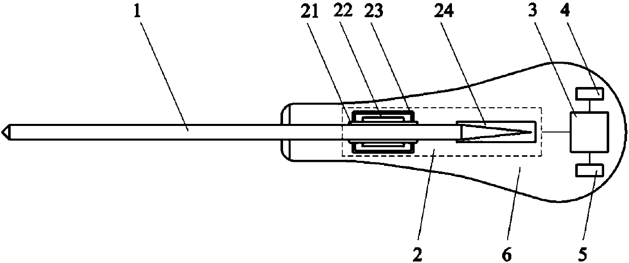
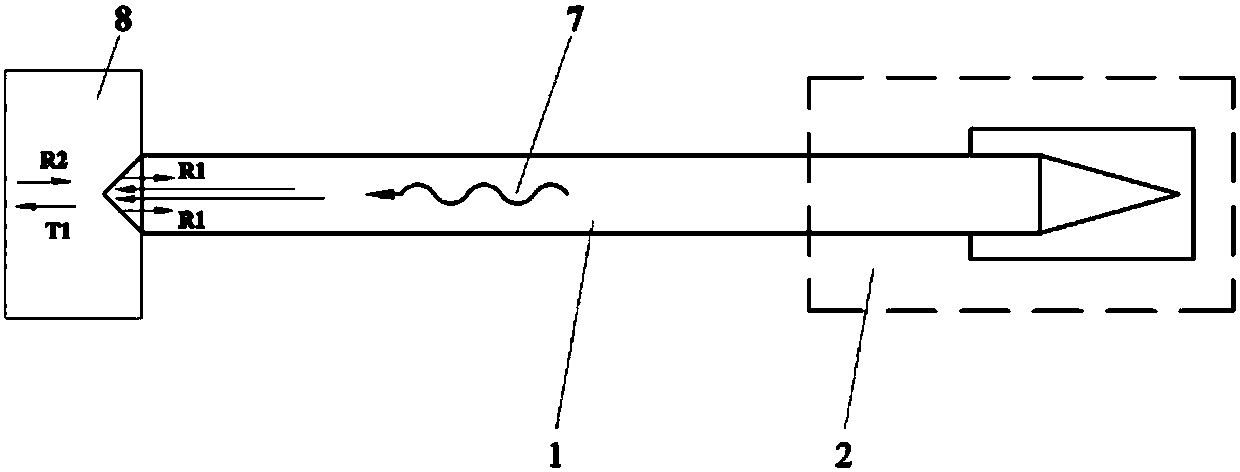
[0052] Such as figure 1 As shown, this embodiment provides an electromagnetic acoustic orthopedic surgery guide device, including a drill pipe 1, an electromagnetic acoustic sensor 2, a measuring controller 3, a warning device 4, and a power supply 5. The drill pipe 1 includes a front end for surgical drilling and The corresponding rear end is used for drilling. The electromagnetic acoustic sensor 2 is arranged at the rear end of the drill pipe 1 to excite and receive ultrasonic waves. The measuring controller 3 is respectively connected to the electromagnetic acoustic sensor 2, the warning devic...


PUM


Abstract

The invention relates to an electromagnetic-acoustic orthopedic surgery guiding device and an alarm method thereof. The device includes a drill rod, an electromagnetic acoustic sensor, a measurement and control instrument, an alarm indicator and a power source. The drill rod comprises a front end for surgery drilling and a corresponding rear end. The electromagnetic acoustic sensor is arranged atthe rear end of the drill rod. The measurement and control instrument is connected with the electromagnetic acoustic sensor, the alarm indicator and the power source separately. The electromagnetic acoustic sensor comprises a metallic sheath, a coil, a permanent magnet and a sound absorption sleeve. The metallic sheath is in interference fit with the drill rod. The coil winds around the metallic sheath. The permanent magnet is connected with the metallic sheath. The sound absorption sleeve is firmly matched with the endmost portion of the drill rod. Compared with the prior art, the electromagnetic-acoustic orthopedic surgery guiding device and the alarm method thereof can overcome the influence of body fluid on detection in an electrical conductivity detection method, precisely measure thethickness of front tissue, and guide spinal surgery drilling.

Description

Technical field [0001] The invention relates to an ultrasonic sensor and medical equipment in the field of biomedical engineering, in particular to an electromagnetic acoustic orthopedic surgery guide device and an alarm method thereof, and more specifically to a device for detecting biological tissues in orthopedic spinal surgery Ultrasonic sensors and equipment to assist drilling. Background technique [0002] At present, there are four types of bone surgery navigation methods at home and abroad: mechanical tool-assisted monitoring screw implantation, intraoperative evoked potential monitoring screw implantation, computer image-assisted monitoring screw implantation and simple auxiliary detection surgical tools. The advantages of the first method are simple and easy to implement, low cost, and good safety; the disadvantage is that it is more dependent on the clinical experience of the doctor. The advantage of the second method is that it can prevent complications such as nerve...

Claims


Application Information

Patent Timeline
IPC IPC(8): A61B17/17
Inventor 雷华明吉小军李红兵
Owner SHANGHAI JIAO TONG UNIV
Why Eureka
Try Eureka