
Detection method and kit for detecting activity of acidic hydrolases in lysosomes

A detection kit and acid hydrolysis technology are applied in the field of detection methods and detection kits for acid hydrolase activity in lysosomes, which can solve the problems of low sensitivity, narrow detection range, weak anti-interference ability and the like, and achieve strong anti-interference ability. , good reproducibility and high sensitivity

Inactive Publication Date: 2018-02-02
SHANGHAI CHROMYSKY MEDICAL RES
View PDF0 Cites 3 Cited by
  • Summary
  • Abstract
  • Description
  • Claims
  • Application Information

AI Technical Summary

Problems solved by technology

At present, fluorescence analysis and colorimetric analysis are mostly used in the detection of hydrolase activity at home and abroad. Most of the colorimetric methods use disodium p-nitrophenylphosphate as the color substrate, because the substrate is catalyzed by alkaline phosphatase. Hydrolysis produces a yellow terminal product with a maximum absorption peak at 405nm, thereby realizing the quantitative colorimetric analysis of alkaline phosphatase activity, but this method has disadvantages such as low sensitivity, narrow detection range and weak anti-interference ability, and the bottom The substance has poor stability, is easy to crystallize, and is easy to decompose under light conditions, resulting in "false positive" results

Method used


Image

  • Detection method and kit for detecting activity of acidic hydrolases in lysosomes
  • Detection method and kit for detecting activity of acidic hydrolases in lysosomes
  • Detection method and kit for detecting activity of acidic hydrolases in lysosomes

Examples


Embodiment Construction

[0046] Below in conjunction with embodiment the present invention is described in further detail.

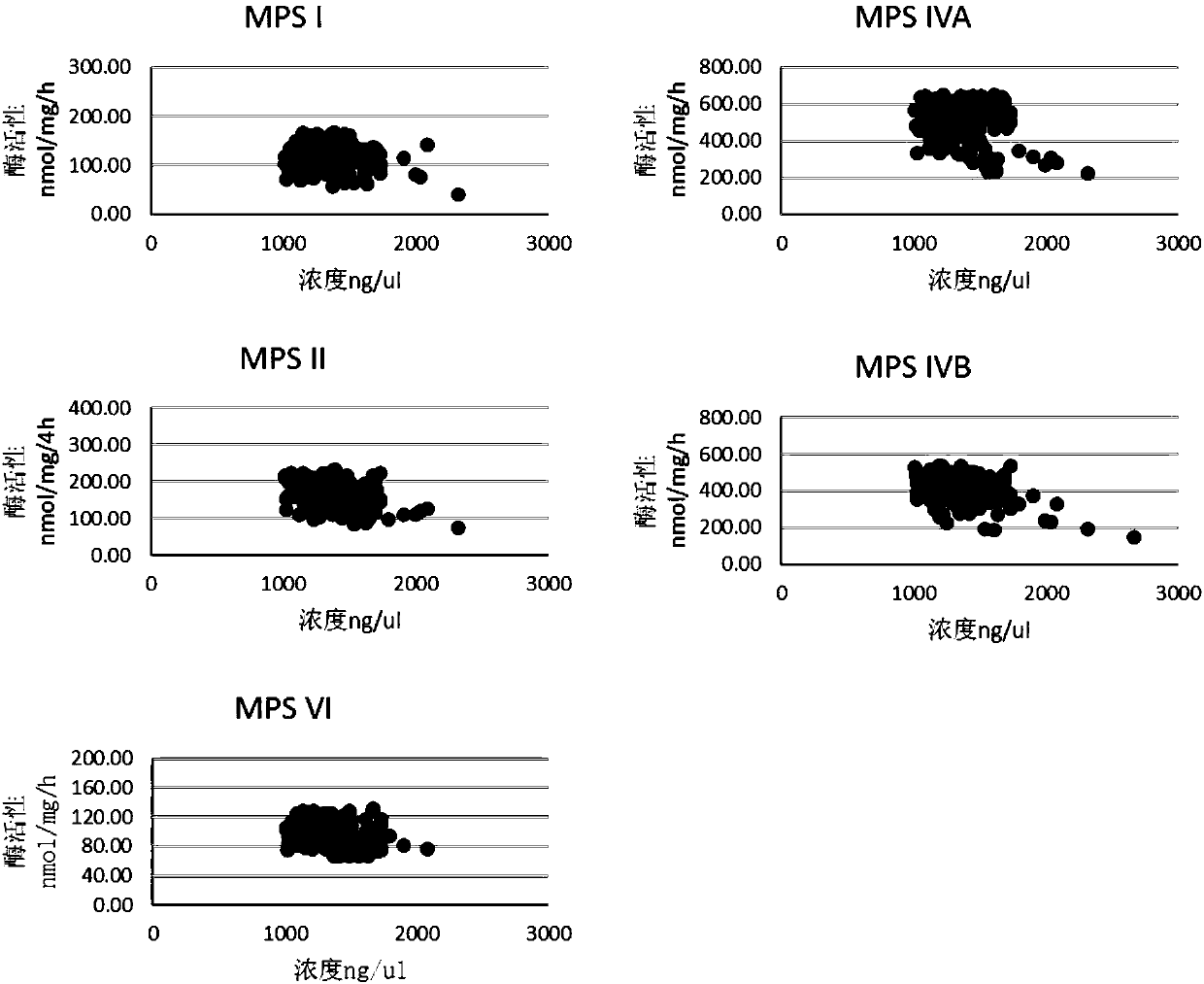
[0047] Take a sample of leukocytes to be tested after extraction, add 200ul of purified water for ultrasonic cell disruption (50W, 1 min in ice bath), and use BCA kit to measure the protein concentration, the concentration of the sample to be tested is 1259ng / ul.

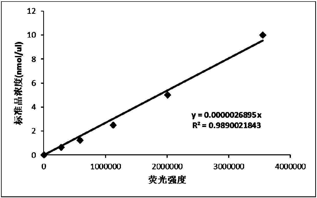
[0048] The hydrolase is α-L-iduronidase, and 10 μl of the substrate, 10 μl of D-glucaric acid-1,4-lactone solution, and the cell homogenate of the sample to be tested are respectively added to the EP tube 10 μl, mix well and put in water bath at 37°C for 1h. After 1 hour of water bath at constant temperature, 200 ul of stop solution was added to terminate the reaction, and the fluorescence intensity was measured by a multi-functional microplate reader. The reaction time is 1 hour, the reaction volume is 230 ul, and the detection volume is 200 ul. The α-L-iduronidase enzyme activity of the sample to be tested is 167.8...


PUM


Abstract

The invention belongs to the technical field of biochemical analysis and detection, in particular to a detection method and a detection kit for acid hydrolase activity in lysosomes. The present invention firstly designs a detection system comprising 5 kinds of mucopolysaccharide acid hydrolase in lysosomes, these five kinds of enzymes are respectively: α-L-iduronidase, iduronate sulfatase, β-N-acetyl half Lactosamine‑6‑sulfatase, β‑galactosidase and arylsulfatase B enzymes. The present invention utilizes the characteristic of fluorescence of 4-methylumbelliferone, uses the new substrate produced by combining the 4-methylumbelliferone group obtained by chemical synthesis with the enzyme substrate, and adopts the acid buffer solution containing the new substrate and mucopolysaccharide The hydrolase reacts, and the free 4-methylumbelliferone fluorescent substance is released after the hydrolase hydrolyzes the substrate. The higher the activity of the hydrolase in the sample, the more the amount of 4-methylumbelliferone released by hydrolyzing the substrate, and the higher the fluorescence intensity generated by the fluorescence detection, so as to realize the quantitative detection of the hydrolase activity. The invention has high detection sensitivity, good selectivity, strong anti-interference ability, good reproducibility and easy operation.

Description

technical field [0001] The invention belongs to the technical field of biochemical analysis and detection, and in particular relates to a detection method and a detection kit for acid hydrolase activity in lysosomes. Background technique [0002] Hydrolase is a general term for a class of enzymes that catalyze hydrolysis reactions. It is widely distributed in the human body and plays an important role in human metabolism and biological regulation and other life activities. Lysosomes are vesicular organelles rich in a variety of high-concentration acid hydrolases in cells, and are important organelles responsible for the decomposition and digestion of various substances in cells. A series of hereditary metabolic diseases caused by the loss of lysosomal enzyme activity are collectively referred to as lysosomal diseases. ). The occurrence of lysosomal diseases is due to the defect of one or more enzymes in lysosomes, resulting in the accumulation of their metabolites in tissu...

Claims


Application Information

Patent Timeline
IPC IPC(8): G01N21/64G01N33/573
CPCG01N21/6428G01N33/573G01N2333/914G01N2333/938
Inventor 赵书民顾学范龚虎涛陈林周巍金云舟
Owner SHANGHAI CHROMYSKY MEDICAL RES
Learn More