Osteochondral tissue engineering scaffold material and preparation method thereof

A tissue engineering scaffold and osteochondral technology, applied in the field of medical materials, can solve the problems of no bionic layered structure and poor biomechanical strength

Active Publication Date: 2020-11-27
SHENZHEN LANDO BIOMATERIALS
View PDF4 Cites 0 Cited by
  • Summary
  • Abstract
  • Description
  • Claims
  • Application Information

AI Technical Summary

Problems solved by technology

[0003] Traditional osteochondral tissue engineering scaffold materials often do not have a biomimetic layered structure, or have a certain biomimetic layered structure but poor biomechanical strength

Method used


Image

  • Osteochondral tissue engineering scaffold material and preparation method thereof
  • Osteochondral tissue engineering scaffold material and preparation method thereof
  • Osteochondral tissue engineering scaffold material and preparation method thereof

Examples


preparation example Construction

[0058] The preparation method of the above-mentioned osteochondral tissue engineering scaffold material of one embodiment, comprises the following steps:

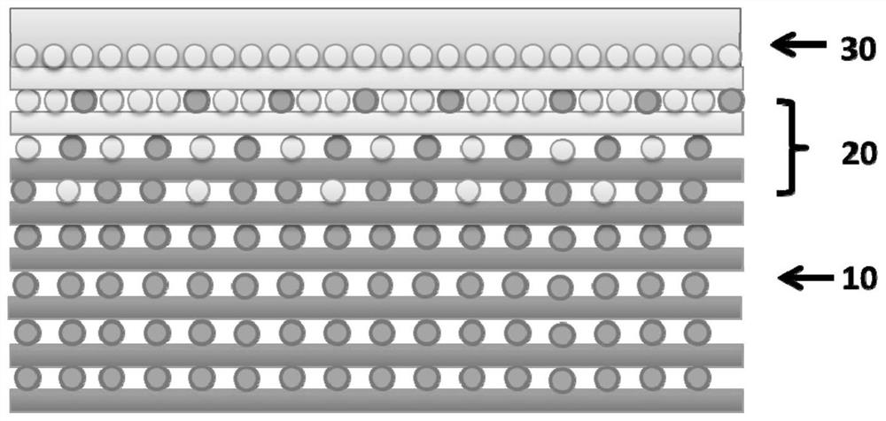
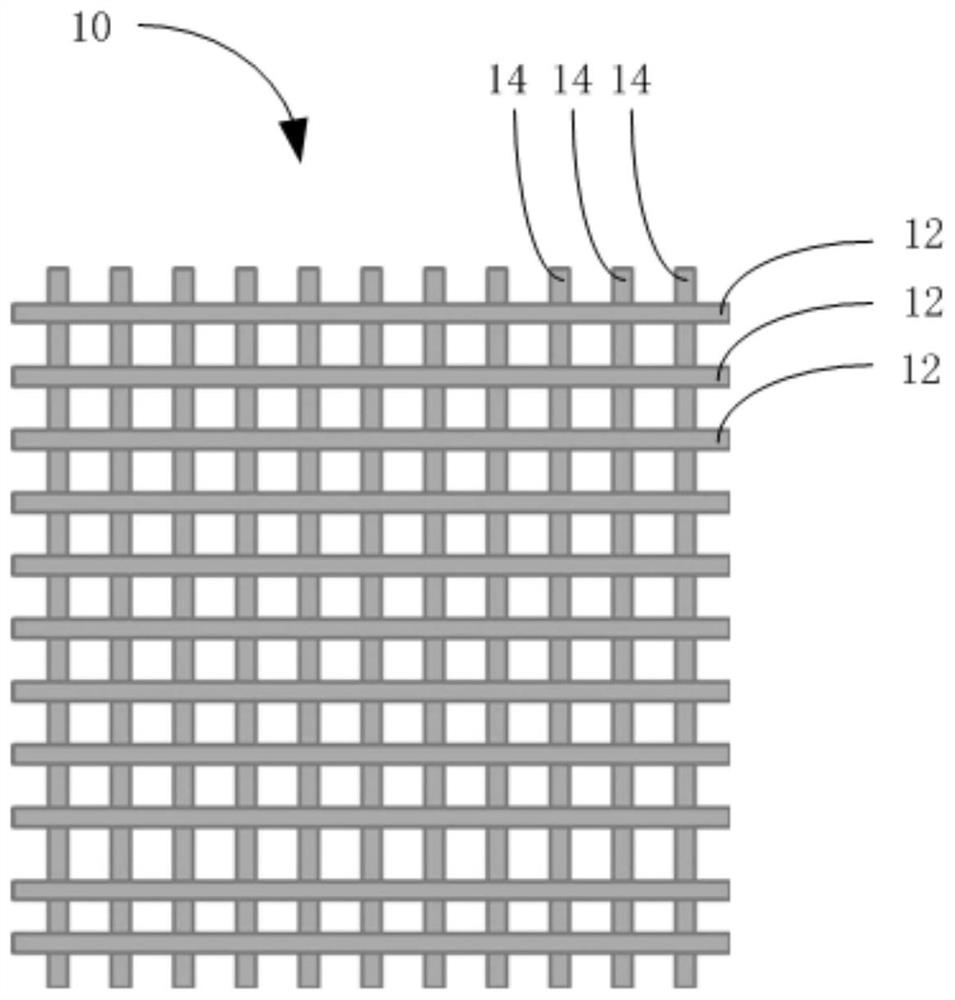
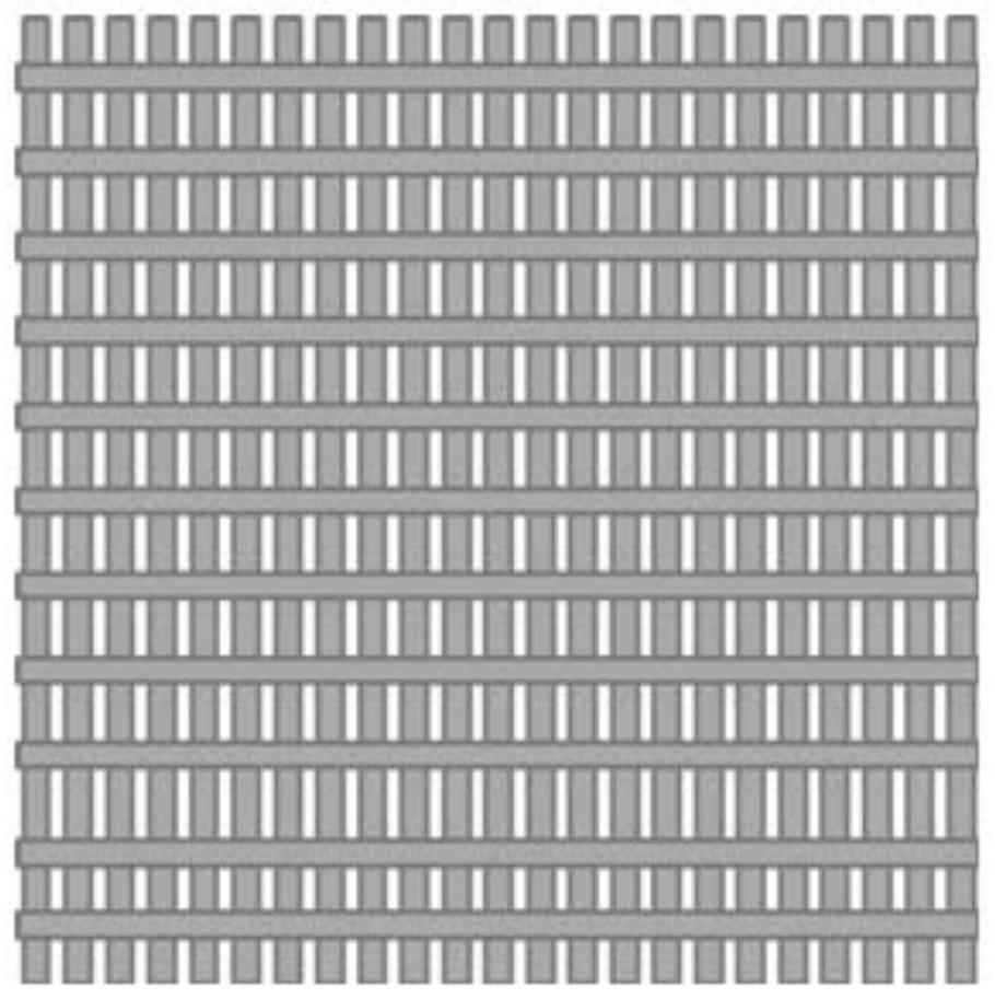
[0059] S10. Using 3D printing technology, input the preset three-dimensional model of the above-mentioned osteochondral tissue engineering scaffold material including subchondral bone layer 10, calcified cartilage layer 20, and cartilage layer 30 that are sequentially stacked and changed gradually, and set the temperature of the printing platform to 10°C ~37°C, the pressure is 0.2Mpa~0.8Mpa.

[0060] S20, adding the polyaryletherketone-based material and the artificial polymer material into the first barrel and the second barrel respectively, selecting a suitable needle, and raising the temperature of the first barrel to the melting point of the polyaryletherketone-based material As mentioned above, the temperature of the second cylinder is raised above the melting point of the artificial polymer material.

[0061] S30 , t...

Embodiment 1

[0078] 1. Input the designed osteochondral tissue engineering scaffold material model into the computer. The osteochondral tissue engineering scaffold material model has a three-layer structure: cartilage layer, calcified cartilage layer and subchondral bone layer.

[0079] 2. Set the printing parameters and operating procedures of the osteochondral tissue engineering scaffold material, and print the osteochondral tissue engineering scaffold material.

[0080] 3. During the printing process, the platform temperature is set to 25°C, and the pressure is set to 0.5MPa.

[0081] 4. Add L-polylactic acid with a molecular weight of 100,000 to the first barrel, select the first needle of 0.15mm, and raise the temperature of the first barrel and the first needle to 190°C. Add polyether ether ketone to the second barrel, select a second needle of 0.15 mm, and increase the temperature of the second barrel and the second needle to 400°C.

[0082] 5. Run the program of the second barrel ...

Embodiment 2

[0086] 1. Input the designed osteochondral tissue engineering scaffold material model into the computer. The osteochondral tissue engineering scaffold material model has a three-layer structure: cartilage layer, calcified cartilage layer and subchondral bone layer.

[0087] 2. Set the printing parameters and operating procedures of the osteochondral tissue engineering scaffold material, and print the osteochondral tissue engineering scaffold material.

[0088] 3. During the printing process, the platform temperature is set to 37°C and the pressure is set to 0.6MPa.

[0089] 4. Add polylactic acid-glycolic acid copolymer with a molecular weight of 100,000 to the first cylinder, select the first needle of 0.20mm, and raise the temperature of the first cylinder and the first needle to 160°C. Add polyether ketone ketone to the second barrel, select a second needle of 0.20 mm, and raise the temperature of the second barrel and the second needle to 390°C.

[0090] 5. Run the progra...


PUM

PropertyMeasurementUnit
diameteraaaaaaaaaa
pore sizeaaaaaaaaaa
diameteraaaaaaaaaa

Abstract

The invention discloses an osteochondral tissue engineering scaffold material and a preparation method thereof. The osteochondral tissue engineering scaffold material comprises a subchondral bone layer, a calcified cartilage layer and a cartilage layer which are sequentially stacked; the subchondral bone layer comprises a first subchondral bone fiber and a second subchondral bone fiber which intersect with each other; the calcified cartilage layer comprises a first calcified cartilage fiber and a second calcified cartilage fiber which intersect with each other; and the material of the cartilage layer is a mixture of type I collagen and chondroitin sulfate. The subchondral bone layer includes the first subchondral bone fiber and the second subchondral bone fiber which intersect with each other, and the calcified cartilage layer includes the first calcified cartilage fiber and the second calcified cartilage fiber which intersect with each other, so the osteochondral tissue engineering scaffold material has a good biomechanical strength and a good application prospect.

Description

technical field [0001] The invention relates to the field of medical materials, in particular to an osteochondral tissue engineering scaffold material and a preparation method thereof. Background technique [0002] Articular cartilage damage caused by arthritis, acromegaly, trauma, metabolic disorders, degenerative diseases, and tumors has become a difficult and hot spot in clinical orthopedics. Articular cartilage lacks structures such as blood vessels, nerves, and lymphatic vessels, and mainly relies on synovial fluid to provide nutrition, so its self-repair ability is limited. Clinical treatment methods for cartilage injuries generally include conservative treatment, joint cavity irrigation, microfracture, perichondrium transplantation, autologous cartilage transplantation, allogeneic articular cartilage transplantation, and total knee replacement. However, these methods cannot completely repair articular cartilage and cannot Obtain satisfactory clinical efficacy (docume...

Claims


Application Information

Patent Timeline
Patent Type & Authority Patents(China)
IPC IPC(8): A61L27/40A61L27/18A61L27/24A61L27/20A61L27/52A61L27/50B33Y10/00B33Y80/00B33Y70/00
CPCA61L27/18A61L27/20A61L27/24A61L27/50A61L27/52A61L2430/06B33Y10/00B33Y70/00B33Y80/00C08L61/16C08L67/04C08L5/08
Inventor 叶晓玲郭远军谭荣伟佘振定
Owner SHENZHEN LANDO BIOMATERIALS
Why Eureka
Try Eureka