

Biosensor for detecting uracil glycosylase (UDG) and application thereof
A uracil glycosylase and biosensor technology, applied in the field of biosensors, can solve the problems of complex operation, high cost, low specificity and sensitivity, etc., and achieve the effects of good repeatability, simple operation and good specificity
- Summary
- Abstract
- Description
- Claims
- Application Information
AI Technical Summary
Problems solved by technology
Method used
Image

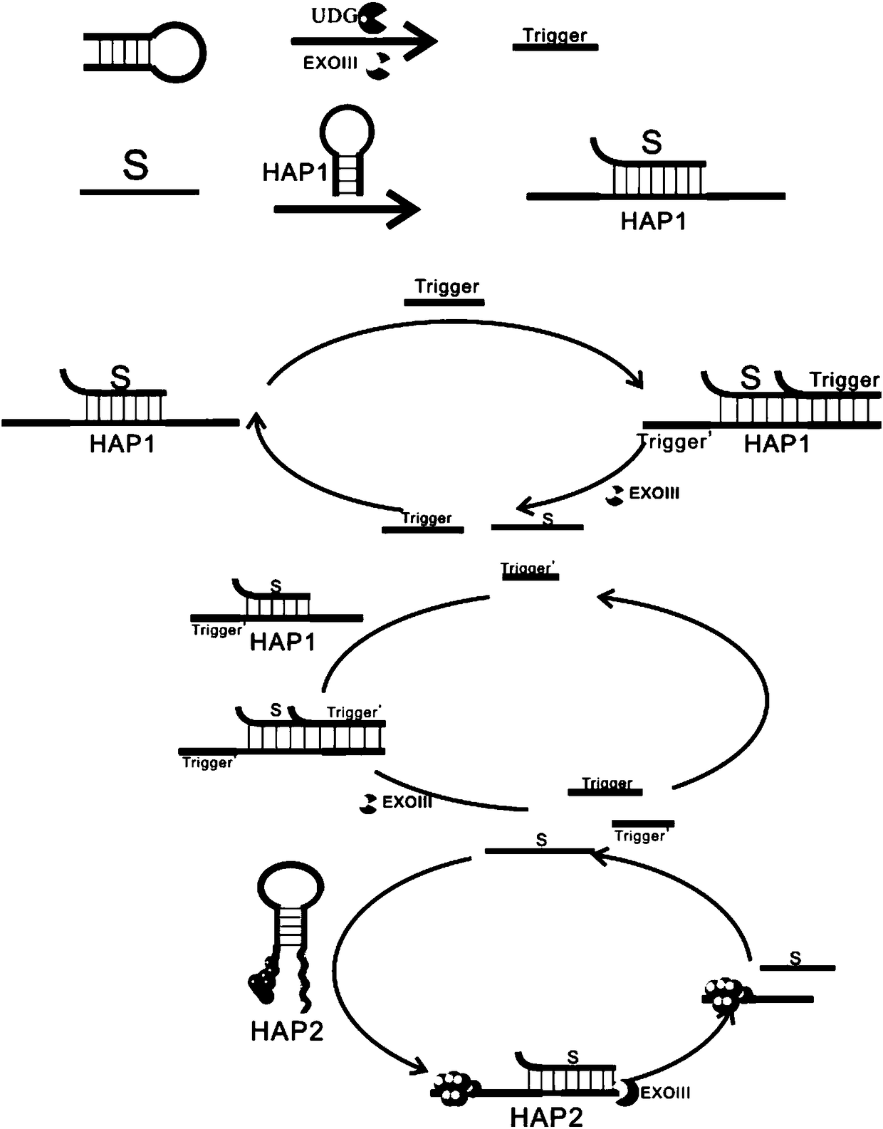
Examples
Embodiment 1
[0033] Example 1 Preparation of HAP2-AgNCs.
[0034] Configure PB buffer solution (concentration is 20mM), PB buffer solution is composed of disodium hydrogen phosphate and sodium dihydrogen phosphate, weigh 0.7163g disodium hydrogen phosphate and 0.3120g sodium dihydrogen phosphate respectively, and make 100ml solution each , and then take a part of disodium hydrogen phosphate and mix a part of sodium dihydrogen phosphate, adjust the pH value of the mixed solution to 6.5 and then set aside.
[0035] Formulating AgNO 3 Concentration of 2mM, volume of 1mL, AgNO 3 Ready to use, store away from light.
[0036] Prepare NaHBO 4 Concentration 2mM, volume 1mL, NaHBO 4 It is ready-to-use and prepared with ice water at 0°C.
[0037] Take a 1 mL centrifuge tube, add 76 μL of PB (20 mM), add 15 μL of HAP2 (100 μM), add 4.5 μL of AgNO 3 (2mM), shake for 1min, put in 4℃ refrigerator for 30min; then add 4.5μL NaHBO 4 (2mM) in the reaction system, shake for 1min, and place in a refrig...
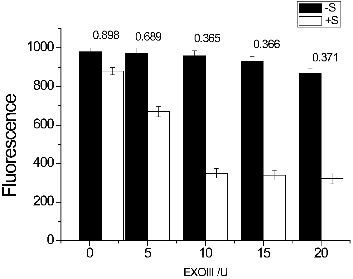
Embodiment 2
[0039] Example 2 The change of fluorescence intensity with the concentration of ExoIII.
[0040] Mix 2 μL of S chain (100 μM), 2 μL of HAP1 (100 μM), and 2 μL of NEBuffer2.1, and let it react at 37°C for 2 hours to obtain S-HAP1 hybrid double strands for use;
[0041] Mix 2 μL of UDG template (1 μM), 3 μL of ExoIII (1U / μL, 5U / μL, 10U / μL, 15U / μL, 20U / μL), 4 μL of NEBuffer 2.1, 2 μL of S-HAP1 hybrid double strand (5 μM) , 8 μL of HAP2-AgNCs (15 μM) and ultrapure water (21 μL) were mixed to measure the fluorescence intensity, then 1 μL of UDG (50 U / mL) was added, reacted at 37 ° C for 2 hours, and the fluorescence intensity was detected;
[0042] The result is as figure 2 As shown, among them, "-S" represents the fluorescence intensity when there is no free S chain in the system, that is, the fluorescence intensity when UDG is not added; "+S" represents the fluorescence intensity when there is a free S chain in the system, That is, the fluorescence intensity after adding UDG, ...
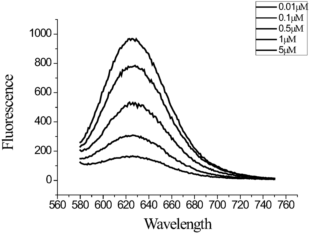
Embodiment 3
[0043] Example 3 Fluorescence intensity changes with the concentration of S-HAP1 hybrid duplex.
[0044]Mix 2 μL of S chain (100 μM), 2 μL of HAP1 (100 μM), and 2 μL of NEBuffer2.1, and let it react at 37°C for 2 hours to obtain S-HAP1 hybrid double strands for use;
[0045] Mix 2 μL of UDG template (1 μM), 3 μL of ExoIII (10 U / μL), 4 μL of NEBuffer 2.1, 2 μL of S-HAP1 hybrid double-stranded (0.01 μM, 0.1 μM, 0.5 μM, 1 μM, 5 μM), 8 μL of HAP2-AgNCs (15 μM) and ultrapure water (21 μL) were mixed, and the fluorescence intensity was measured, then 1 μL of UDG (50 U / mL) was added, reacted at 37°C for 2 hours, and the fluorescence intensity was detected.
[0046] The result is as image 3 , the detected fluorescence signal intensity gradually decreases with the concentration of S-HAP1 hybrid duplex in the range of 0.01-5 μM. When the concentration of S-HAP1 hybrid duplex in the reaction system is 5 μM, the fluorescence intensity value is large.
PUM
 Abstract
Description
Claims
Application Information