Method for isolating target cells from a blood sample and use thereof

A blood sample and target cell technology, applied in the field of separating target cells, can solve the problems that need to be strengthened, the detection of specific gene loci is difficult, incomplete, etc.

Active Publication Date: 2022-02-01
SHENZHEN HUADA GENE INST
View PDF7 Cites 0 Cited by
  • Summary
  • Abstract
  • Description
  • Claims
  • Application Information

AI Technical Summary

Problems solved by technology

However, the use of fetal cell-free DNA has its own defects. First, in addition to fetal cell-free DNA, there are also a large amount of maternal DNA in maternal blood; at the same time, fetal cell-free DNA in maternal blood is fragmented and exists as an incomplete genome. , so at present it is mainly aimed at the detection of abnormal chromosome number, and there are great difficulties in the detection of specific gene loci
[0005] Therefore, the current research on the detection of fetal DNA in the peripheral blood of pregnant women still needs to be strengthened

Method used


Image

  • Method for isolating target cells from a blood sample and use thereof
  • Method for isolating target cells from a blood sample and use thereof
  • Method for isolating target cells from a blood sample and use thereof

Examples


Embodiment 1

[0092] Example 1 Pretreatment of Peripheral Blood of Pregnant Women and Sorting of Single Cells

[0093] In this embodiment, a pregnant woman sample from a normal male fetus is used, sample number: CFCCYY; male fetus; blood volume: 10 mL; gestational week: 13 weeks.

[0094] Take 10mL of pregnant women's peripheral blood with a heparin tube, add the blood sample to 100mL BD split red solution diluted to 1X in advance, mix gently, and split red at room temperature until the blood is clear, centrifuge to absorb the supernatant, Gently pipette the cell pellet, wash twice with PBS containing 2% FBS, resuspend the cell pellet in 80 microliters of PBS containing 2% FBS, and blow evenly. Add CD45 antibody magnetic beads, incubate at 4°C for 15 minutes, centrifuge at 300g for 10 minutes, discard the supernatant, and resuspend the pellet in PBS containing 2% FBS. Assemble a magnetic bead sorting system (Miltenyi), collect CD45-negative outflow cells, centrifuge to remove the supernata...

Embodiment 2

[0095] Whole genome amplification of embodiment 2 single cell

[0096] The single cells obtained in Example 1 were subjected to whole genome amplification, as follows:

[0097] 1. Preparation of kits for single-cell whole-genome amplification

[0098] The kit consists of cell denaturation lysate, neutralization buffer and amplification system.

[0099] Cell denaturation lysate: composed of solute and solvent; solvent is water; solute and its concentration are as follows: 0.1mM KOH, 1mM EDTA and 0.1M DTT.

[0100] Neutralization buffer: composed of solute and solvent; solvent is water; solute and its concentration are as follows: 60mM KH 2 PO 4 and 5mMK 2 HPO 4 .

[0101] The amplification system includes: 10×Phi29buffer, water, 1.8-2.2×10 -8 mol dNTP, total concentration of primers is 1.8-2.2×10 -11 mol N8primers, 4.5-5.5×10 -3 mg BSA, 0.0045-0.0055 μL Pluronic F68 and 9-11UPhi29 DNA polymerase. Specifically, taking 40.3 μL as an example, the amplification system: 30...

Embodiment 3

[0113] Example 3 Q-PCR detection and STR site detection of single cells

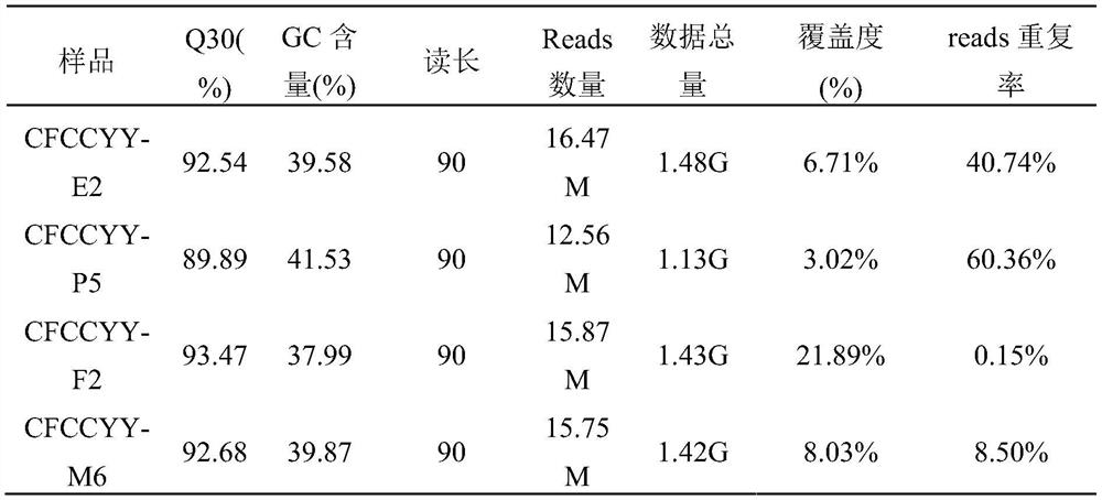
[0114] The 114 single-cell amplification products obtained from the above amplification were used Trio DNAQuantification Kit (Applied Biosystems TM ) for Q-PCR detection. The instrument adopts ViiA 7 real-time fluorescent quantitative PCR instrument, and the reaction conditions of amplification refer to the procedures provided by the kit. Among the 114 products, the amplification products of 4 cells had obvious Y amplification signal.

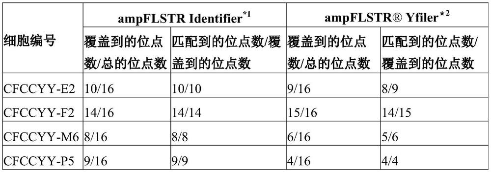
[0115] The inventor further used the ampFLSTRIdentifier PCR amplification kit and the genomes of these 4 single-cell amplification products and parents The Yfiler amplification kit was used for STR detection, and the results are shown in Table 1:

[0116] Table 1: Conventional STR and Y chromosome STR loci

[0117]

[0118] Note: * 1: The ampFLSTR Identifier PCR Amplification Kit can simultaneously amplify and detect 16 STR sites at the same time, so the total ...


PUM


Abstract

Provided are a method for obtaining target cells from a blood sample and uses thereof, the method comprising: (1) subjecting the blood sample to immunomagnetic bead sorting, so as to obtain a first sorting product containing the target cells; The first sorted product is subjected to flow cytometric sorting to obtain a second sorted product containing the target cell; and (3) isolating the second sorted product using a pipette method so as to obtain the target in the form of a single cell cell.

Description

[0001] priority information [0002] none. technical field [0003] The invention relates to the field of biotechnology, in particular, the invention relates to a method for isolating target cells from a blood sample and an application thereof. Background technique [0004] In recent years, with the development of high-throughput sequencing technology, the use of high-throughput sequencing methods to detect fetal cell-free DNA in the peripheral blood of pregnant women to determine the abnormal number of fetal chromosomes has become more and more widely recognized. However, the use of fetal cell-free DNA has its own defects. First, in addition to fetal cell-free DNA, there are also a large amount of maternal DNA in maternal blood; at the same time, fetal cell-free DNA in maternal blood is fragmented and exists as an incomplete genome. , so at present it is mainly aimed at the detection of abnormal chromosome number, and there are great difficulties in the detection of specif...

Claims


Application Information

Patent Timeline
Patent Type & Authority Patents(China)
IPC IPC(8): C12N5/078C12N5/073C12N15/10C12M1/42C12Q1/686C12Q1/6869G01N33/53
CPCC12N15/1093C12M47/04C12Q1/6806G01N33/54326C12Q2545/10C12Q2565/101C12Q2565/626
Inventor 朱珠刘萍顾颖康雄斌夏军邱咏
Owner SHENZHEN HUADA GENE INST
Why Eureka
Try Eureka