Short-T2-effect endorectal/endovaginal magnetic resonance imaging contrast agent and preparation method thereof

A magnetic resonance imaging, vaginal cavity technology, applied in the direction of magnetic resonance/magnetic resonance imaging contrast agent, echo/ultrasonic imaging agent, etc. couplant confusion and other problems, to achieve the effect of shortening the relaxation time, reducing the signal intensity, and long shelf life

Active Publication Date: 2018-09-11
SHENZHEN PEOPLES HOSPITAL
View PDF1 Cites 0 Cited by
  • Summary
  • Abstract
  • Description
  • Claims
  • Application Information

AI Technical Summary

Problems solved by technology

[0004] At present, the filling agent commonly used in clinical MRI examination and diagnosis of rectum and vagina is ultrasonic coupling agent. Cavity organs can eliminate the influence of cavity and intestinal contents to improve the display of lesions. It is helpful for MRI examination and diagnosis of rectum and vagina. Compared with water as a filling agent, ultrasonic coupling agent has no effect. Fluidity, can be properly molded, does not require a specific container, can be directly filled in the rectum or vagina; long shelf life, convenient storage, but, in the magnetic resonance imaging, the ultrasonic coupling agent is high signal on T2-weighted imaging, which directly It affects the display of lesions, such as colorectal cancer, especially in diffusion-weighted images (Diffusion-weighted Image, referred to as DWI), which will affect the T2 penetration effect, making the ultrasound coupling agent confused with the diffusion-limited human tissue lesion mass, This seriously affects the MRI examination and diagnosis of the rectum and vagina, and ultimately affects the treatment effect. At present, in clinical practice, an ideal MRI contrast agent for the rectum and vagina that does not confuse the display of lesions is still in a vacancy, so , a magnetic resonance imaging contrast agent that can clearly display rectal and vaginal lesions is an urgent need for clinical and scientific research

Method used


Image

  • Short-T2-effect endorectal/endovaginal magnetic resonance imaging contrast agent and preparation method thereof
  • Short-T2-effect endorectal/endovaginal magnetic resonance imaging contrast agent and preparation method thereof
  • Short-T2-effect endorectal/endovaginal magnetic resonance imaging contrast agent and preparation method thereof

Examples


preparation example Construction

[0036] In this embodiment, the preparation method of the above-mentioned contrast agent includes the steps of: taking the above-mentioned iron dextran, ultrasonic coupling agent and water of specified mass respectively; mixing the above-mentioned iron dextran and water at a specified temperature and putting them into the above-mentioned ultrasonic coupling agent and stirring Uniform; placed in an ultrasonic degassing tank for degassing treatment; radiation disinfection treatment by cobalt 60, since the above-mentioned contrast agent is used in the rectal and vaginal cavities of the human body in magnetic resonance imaging, care should be taken during the preparation process Safety, hygiene and disinfection issues, so the preparation environment should be a sterile room; after the above-mentioned iron dextran solution is placed in the above-mentioned ultrasonic coupling agent, the stirring method includes rotating stirring and stirring up and down. Artifacts will be produced dur...

specific Embodiment 1

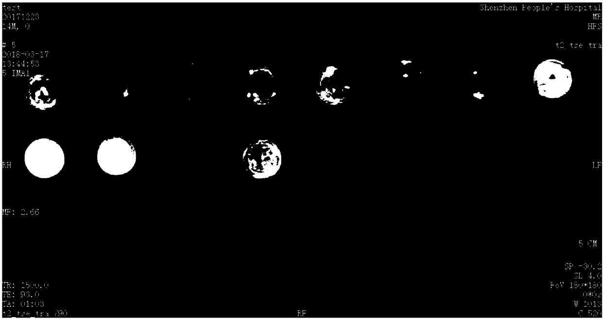
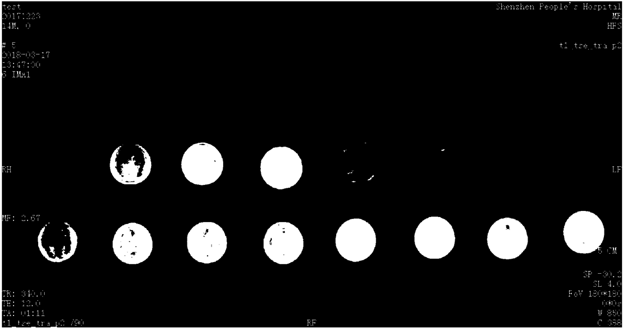
[0042] As shown in Table 2, take 10g of the above-mentioned ultrasonic coupling agent and pour it into the above-mentioned test tube, which is recorded as A1, and take 10g of the above-mentioned iron dextran solution and pour it into the above-mentioned test tube, which is recorded as B1, and take 0.2g of the above-mentioned iron dextran solution and put it in 9.8g of the above-mentioned ultrasonic coupling agent , stirred evenly, poured into the above-mentioned test tube, sterilized with the above-mentioned cobalt 60 radiation after degassing through the above-mentioned ultrasonic degassing tank, and obtained the above-mentioned contrast agent with a total weight of 10 g, an iron content of 1.25 mg, and an iron content of 0.125 mg / g. Record it as C1, put the above A1, B1 and C1 under the above 3.0 TMRI for scanning, and repeat this test 3 to 10 times.

[0043]

A1

B1

C1

Iron dextran solution (g)

0

10

0.2

Contains about iron (mg)

0

...

specific Embodiment 2

[0046] As shown in Table 3, take 10g of the above-mentioned ultrasonic coupling agent and pour it into the above-mentioned test tube, which is recorded as A2, and take 10g of the above-mentioned iron dextran solution and pour it into the above-mentioned test tube, which is recorded as B2, and take 0.6g of the above-mentioned agent a and place it in 9.4g of the above-mentioned ultrasonic coupling agent. After stirring evenly, pour it into the above-mentioned test tube, and after degassing in the above-mentioned ultrasonic degassing tank, sterilize it with the above-mentioned cobalt 60 radiation to obtain the above-mentioned contrast agent with a total weight of 10 g, an iron content of 3.75 mg, and an iron content of 0.375 mg / g. For C2, the above-mentioned A2, B2 and C2 are scanned under the above-mentioned 3.0 TMRI, and this test is repeated 3 to 10 times.

[0047]

A2

B2

C2

Iron dextran solution (g)

0

10

0.6

Contains about iron (mg)

...


PUM


Abstract

The invention discloses a short-T2-effect endorectal / endovaginal magnetic resonance imaging contrast agent and a preparation method thereof. The short-T2-effect endorectal / endovaginal magnetic resonance imaging contrast agent comprises iron dextran, an ultrasonic coupling agent and water. The short-T2-effect endorectal / endovaginal magnetic resonance imaging contrast agent has the beneficial effects that the iron dextran, the ultrasonic coupling agent and the water are mixed according to a specified proportion to prepare the contrast agent, so that the T2 relaxation time of the ultrasonic coupling agent during magnetic resonance imaging is shortened, the signal strength of the ultrasonic coupling agent on a T2-weighted imaging is reduced, and interference of the ultrasonic coupling agent tolesion display during endorectal / endovaginal magnetic resonance imaging is reduced; the contrast agent has attachment performance with cavity organs such as the rectum / vagina, is a non-liquid colloid, can be properly formed and is not required to be contained in a specific container, the cavity organs such as the rectum / vagina can be well filled, and the rectum or the vagina can be directly filled with the contrast agent; each component is stable, and the contrast agent is long in shelf life and convenient to store.

Description

technical field [0001] The invention relates to the field of medical preparations, in particular to a short T2 effect rectal / vaginal cavity magnetic resonance imaging contrast agent and a preparation method thereof. Background technique [0002] Magnetic Resonance Imaging (MRI for short) is an imaging technique that uses the signal generated by the resonance of atomic nuclei in a magnetic field to reconstruct the image. As a new medical imaging diagnosis technology, it has developed rapidly in recent years. The amount of information provided by magnetic resonance imaging is not only more than that of many other imaging techniques, but also has great potential advantages in diagnosing diseases with the unique information it provides. In magnetic resonance imaging, good image quality is the basis of all clinical diagnosis and research. Therefore, high-quality images are the primary condition for doctors to correctly study, judge, diagnose and treat patients' lesions. [0003]...

Claims


Application Information

Patent Timeline
Patent Type & Authority Applications(China)
IPC IPC(8): A61K49/22A61K49/06
CPCA61K49/06A61K49/22
Inventor 龚静山唐雪贾桂静焦月新周宙冯霞汪娟易芹芹
Owner SHENZHEN PEOPLES HOSPITAL
Why Eureka
Try Eureka