Eureka AIR delivers breakthrough ideas for toughest innovation challenges, trusted by R&D personnel around the world.

Method and device for detecting tumor cell in blood by using stimulated Raman spectrum

A tumor cell and stimulated Raman technology, applied in the field of bioinformatics, can solve the problems of cumbersome blood sample processing, low sensitivity, complicated reagent labeling and operation, and achieve short signal acquisition time, improved sensitivity, and high Raman Effect of Signal Intensity or Brightness

Pending Publication Date: 2018-11-13
上海镭立激光科技有限公司
View PDF0 Cites 4 Cited by
  • Summary
  • Abstract
  • Description
  • Claims
  • Application Information

AI Technical Summary

Problems solved by technology

[0006] The purpose of the present invention is to provide a method and device for detecting tumor cells in blood by using stimulated Raman spectroscopy. The method and device for detecting tumor cells in blood should solve the problem of detecting tumor cells in the prior art. The sensitivity of the method is not high, the processing of blood samples is cumbersome, the need for other reagents to label and the technical problems of complex operation

Method used


Image

  • Method and device for detecting tumor cell in blood by using stimulated Raman spectrum

Examples


Embodiment 1

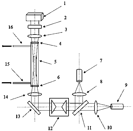
[0024] like figure 1 As shown, the present invention provides a device for detecting tumor cells in blood by using stimulated Raman spectroscopy, comprising a capillary glass tube 5, the two ends of the capillary glass tube 5 are respectively provided with a first optical window 4 and a second optical window 6;

[0025] It also includes a pump laser source 9, and a second collimating lens 10, a beam splitter 11, a pair of axicon mirrors 12 and a reflector 13 are sequentially arranged on the optical path of the pump laser source 9. An objective lens 14 is provided between the reflecting mirror 13 and the second optical window 6; the beam splitter 11 and the laser light path emitted by the pump laser source 9 are at an angle of 45 degrees. , an angle of 90 degrees is formed between the reflecting mirror 13 and the beam splitter 11, and the reflecting mirror 13 reflects the light beam of the pump laser source 9 into the capillary glass tube 5;

[0026] Also includes a Stokes la...

Embodiment 2

[0031]Using the device shown in Example 1 of the present invention, the present invention performs stimulated Raman spectroscopic analysis on lipid droplets in blood. Lipid droplets are composed of a monolayer of phospholipids and a hydrophobic core composed of neutral lipids, and there are many proteins distributed on the surface. Is an "inert" cell inclusion. We take the capillary glass tube length 2mm, internal diameter: 100um. We take 5 ml of blood sample and pump it into the capillary glass tube to circulate, and adjust the Raman scattered light transmission to 2,850 cm -1 the CH 2 Symmetrical vibration for stimulated Raman imaging of lipid droplets. The Stokes laser source 7 and the pump laser source 9 generate two synchronized laser beams with a repetition rate of 80 MHz. Among them, the Stokes laser 7 has a fixed wavelength of 1,040 nm. The pump laser 9 has a tunable wavelength of 680 to 1,300 nm. The beams are then spatially combined by a beam splitter 11 . Tw...


PUM

PropertyMeasurementUnit
lengthaaaaaaaaaa
diameteraaaaaaaaaa

Abstract

The invention discloses a method for detecting a tumor cell in blood by using a stimulated Raman spectrum. The method comprises the following steps: creating a stimulated Raman spectrum database for chemical ingredients of related substances in various related tumor cells, testing stimulated Raman characteristic spectrum data of the characteristic chemical ingredients of various related tumor cells of a test sample, using an intrinsic molecular vibration Raman spectrum signal of the characteristic chemical ingredient of the tumor cell as a marker for stimulated Raman imaging, measuring the chemical substance of the test sample through a Bessel beam based stimulated Raman projection real-time imaging, acquiring information of the characteristic chemical ingredient of the tumor cell in the whole test sample, comparing the stimulated Raman data in the database, and determining whether the test sample contains certain types of tumor cells. The invention further provides a device for detecting the tumor cell in the blood by using the stimulated Raman spectrum. The circulating tumor cell in the blood is detected through the Bessel beam based stimulated Raman projection real-time imaging,and the different tumor cells are detected at high sensitivity.

Description

technical field [0001] The invention belongs to the field of bioinformatics, and relates to a method for detecting tumor cells, in particular to a method and a device for detecting tumor cells in blood by using stimulated Raman spectroscopy. Background technique [0002] Distant metastasis of malignant tumors is one of the main reasons for clinical treatment failure or recurrence of solid malignant tumors. The existence of circulating tumor cells is the root cause of distant metastasis of solid malignant tumors. Circulating tumor cells are tumor cells present in the blood with the antigenic and molecular genetic properties of the original tumor. Circulating tumor cells originate from primary tumors or metastases, and when they encounter the stromal environment of suitable organs and tissues, they will settle and grow here, that is, tumor metastasis occurs. However, most of the tumor cells entering the circulation will lose their activity, and only less than 0.01% can reach...

Claims


Application Information

Patent Timeline
Patent Type & Authority Applications(China)
IPC IPC(8): G01N21/65
CPCG01N21/65
Inventor 龚辉姜辛倪勤
Owner 上海镭立激光科技有限公司
Why Eureka
Eureka Blog
Learn More