Single domain heavy chain antibody for detecting porcine epidemic diarrhea virus, preparation method and application

A porcine epidemic diarrhea and single-domain heavy chain antibody technology, applied in the field of genetic engineering, can solve the problem of few epitopes of PEDVS protein, and achieve good antibody stability and specificity

Active Publication Date: 2021-09-21
INNER MONGOLIA AGRICULTURAL UNIVERSITY
View PDF3 Cites 0 Cited by
  • Summary
  • Abstract
  • Description
  • Claims
  • Application Information

AI Technical Summary

Problems solved by technology

[0006] At present, there are few studies on the epitope of PEDV S protein, and the epitope can stimulate the body to produce antibodies and protect the body from pathogens

Method used


Image

  • Single domain heavy chain antibody for detecting porcine epidemic diarrhea virus, preparation method and application
  • Single domain heavy chain antibody for detecting porcine epidemic diarrhea virus, preparation method and application
  • Single domain heavy chain antibody for detecting porcine epidemic diarrhea virus, preparation method and application

Examples


Embodiment 1

[0026] Example 1 (Construction of a PEDV-specific single domain heavy chain antibody library)

[0027] (1) Mix PEDV virus with Freund's adjuvant (Sigma), immunize one hump of Alxa Bactrian camel, immunize once every 10 days, and immunize 3 times in total. Freund's complete adjuvant is used for the first time, and Freund's complete adjuvant is used for the remaining two times. Incomplete adjuvant, the basic serum was collected before the first immunization, and the serum was collected one week after the last immunization to determine the antibody titer;

[0028] (2) After the 3 times of immunization, extract 150 ml of camel peripheral blood lymphocytes and extract total RNA;

[0029] (3) Reverse transcribe the total RNA into cDNA by RT-PCR;

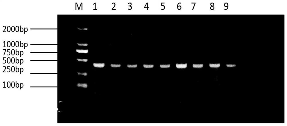
[0030] (4) Using nested PCR to amplify the VHH chain, the size of the fragment is about 400 bp; figure 1 It is the detection electrophoresis diagram of the VHH gene fragment obtained by PCR amplification;

[0031] (5) Using restriction ...

Embodiment 2

[0033] Example 2 (screening PEDV single domain heavy chain antibody)

[0034] (1) Add 2 ml of transformed TG1 cells into 100 ml of 2×TY-AG medium, and incubate at 37°C for 2 hours;

[0035] (2) Add helper phage M13K07 for infection, the infection ratio is M13K07:TG1=20:1, and stand at room temperature for 30 minutes;

[0036] (3) Centrifuge for 10 minutes, resuspend the pelleted cells into 100 ml of 2×YT-AK medium, and culture overnight at 37°C;

[0037] (4) Centrifuge the culture solution at 2200 g for 30 minutes, take the supernatant, precipitate the amplified phage with PEG / NaCl, dissolve the precipitated phage in PBS solution, filter through a 0.22 micron filter, and store at 4°C for later use;

[0038] (5) Dilute the PEDV S protein with coating buffer and couple to the microtiter plate, and place it overnight at 4°C;

[0039] (6) Add 300 microliters of blocking solution to each well on the second day, and block at room temperature for 2 hours;

[0040] (7) Mix the phag...

Embodiment 3

[0043] Example 3 (screening PEDV S protein-specific clones by phage-ELISA method)

[0044] (1) Randomly pick 96 single clones from the cell culture dishes containing phages after the above three rounds of selection, inoculate them in 1 ml of 2×TY-AG medium, and culture overnight at 37°C;

[0045] (2) Transfer the overnight cultured bacterial solution into 1.3 ml 2×YT-AG medium and 10 9In four 24-well plates of M13K07, culture at 150 rpm at 37°C for 2 h, centrifuge at 1000 g for 20 min, resuspend the pellet with 1 ml of 2×YT-AK medium, culture at 250 rpm at 30°C overnight, and collect phage by centrifugation the next day supernatant;

[0046] (3) Mix the above-mentioned recombinant phage with blocking solution 1:1, add to a 96-well microtiter plate coated with 10 μg / ml PEDVS protein, incubate at 37°C for 1 h, add 100 μl to the negative control well 10 9 M13K07;

[0047] (4) Wash away unbound phage with PBST, 100 microliters of 1:5000 diluted HRP / anti-M13 monoclonal antibody...


PUM


Abstract

The invention discloses a single-domain heavy chain antibody for detecting porcine epidemic diarrhea virus, a preparation method and application. The present invention adopts porcine epidemic diarrhea virus to immunize Alxa Bactrian camels, uses its peripheral blood lymphocytes to establish a single-domain heavy chain antibody library against porcine epidemic diarrhea virus, and couples the expressed and purified porcine epidemic diarrhea virus S protein to As an antigen on the enzyme plate plate, the single domain heavy chain antibody library was screened immunologically by using phage display technology, and the single domain heavy chain antibody gene against the S protein of porcine epidemic diarrhea virus was obtained, and the single domain heavy chain antibody gene was transferred into the large intestine In Bacillus, a single-domain heavy-chain antibody strain highly expressed in Escherichia coli was established, and the gene sequence of each single-domain heavy-chain antibody strain was analyzed to obtain the amino acid of the VHH chain of the single-domain heavy-chain antibody against the S protein of porcine epidemic diarrhea virus Sequence SEQ ID NO.8. The antibody of the invention has good stability and specificity, and can be used for detecting and treating PEDV.

Description

technical field [0001] The invention relates to the technical field of genetic engineering, in particular to a single domain heavy chain antibody for detecting porcine epidemic diarrhea virus, a preparation method and application. Background technique [0002] Porcine epidemic diarrhea is caused by porcine epidemic diarrhea virus (Porcine epidemic diarrhea virus, PEDV), a highly contagious intestinal infectious disease characterized by diarrhea, vomiting, dehydration and high lethality to young piglets. [0003] The pathological changes of pigs infected with PEDV are mainly manifested in the atrophy and shedding of intestinal villi in the jejunum and ileum of pigs, which is very harmful to pigs and brings serious impact and economic losses to the pig industry. [0004] The S protein is an important structural protein of PEDV. It is a spike glycoprotein located on the surface of the virus particle. It plays an important biological role in the process of mediating the producti...

Claims


Application Information

Patent Timeline
Patent Type & Authority Patents(China)
IPC IPC(8): C07K16/10G01N33/569A61K39/42A61P31/14
CPCA61P31/14C07K16/005C07K16/10C07K2317/22C07K2317/569C07K2317/76G01N33/56983G01N2333/165
Inventor 包福祥王莉鑫杜雅楠芦婷阿米娜赵鑫鑫关平原
Owner INNER MONGOLIA AGRICULTURAL UNIVERSITY
Why Eureka
Try Eureka