Whole breast tumor small-interval subserial pathological slice preparation method

A production method and slicing technology, which is applied in the field of clinical medical experiment research and medical teaching, can solve the problems such as difficulty in realizing sub-continuous large slicing technology of whole breast, unsatisfactory slicing results, etc., and achieve easy slicing production process and excellent production efficiency , Reduction of residue and toxicity

Active Publication Date: 2018-12-14
WEST CHINA HOSPITAL SICHUAN UNIV
View PDF4 Cites 15 Cited by
  • Summary
  • Abstract
  • Description
  • Claims
  • Application Information

AI Technical Summary

Problems solved by technology

[0011] The purpose of the present invention is to overcome the problems in the prior art that only small-scale pathological tissue slices can be made, the technique of whole breast sub-continuous large slices is difficult to realize, and the slice results are not ideal, and to provide a method for making whole breast sub-continuous large slices

Method used


Image

  • Whole breast tumor small-interval subserial pathological slice preparation method
  • Whole breast tumor small-interval subserial pathological slice preparation method
  • Whole breast tumor small-interval subserial pathological slice preparation method

Examples


Embodiment 1

[0070] Collect tumor specimens from patients with invasive ductal carcinoma of the breast, cut off the obvious excess fat and normal glandular tissue (obviously larger fat after the tumor specimen was obtained, etc.), keep the key parts of the tumor specimen as much as possible, and remove the irrelevant parts. The experience of clinicians can be implemented). Put the tumor specimens in a -80°C refrigerator for 30 minutes, take them out and cut them in parallel at intervals of 15 mm to obtain parallel cut specimen blocks, and mark the position of each cutting edge seam (determine the direction of each specimen and the direction before cutting). arrangement position). Place a piece of gauze on each parallel cut section of the cut specimen block; then, soak and fix it in 4% neutral formaldehyde solution for 24 hours, and the volume of neutral formaldehyde solution is about 22 times the sample volume.

[0071] The formula of neutral formaldehyde solution is as follows: 100 ml of...

Embodiment 2

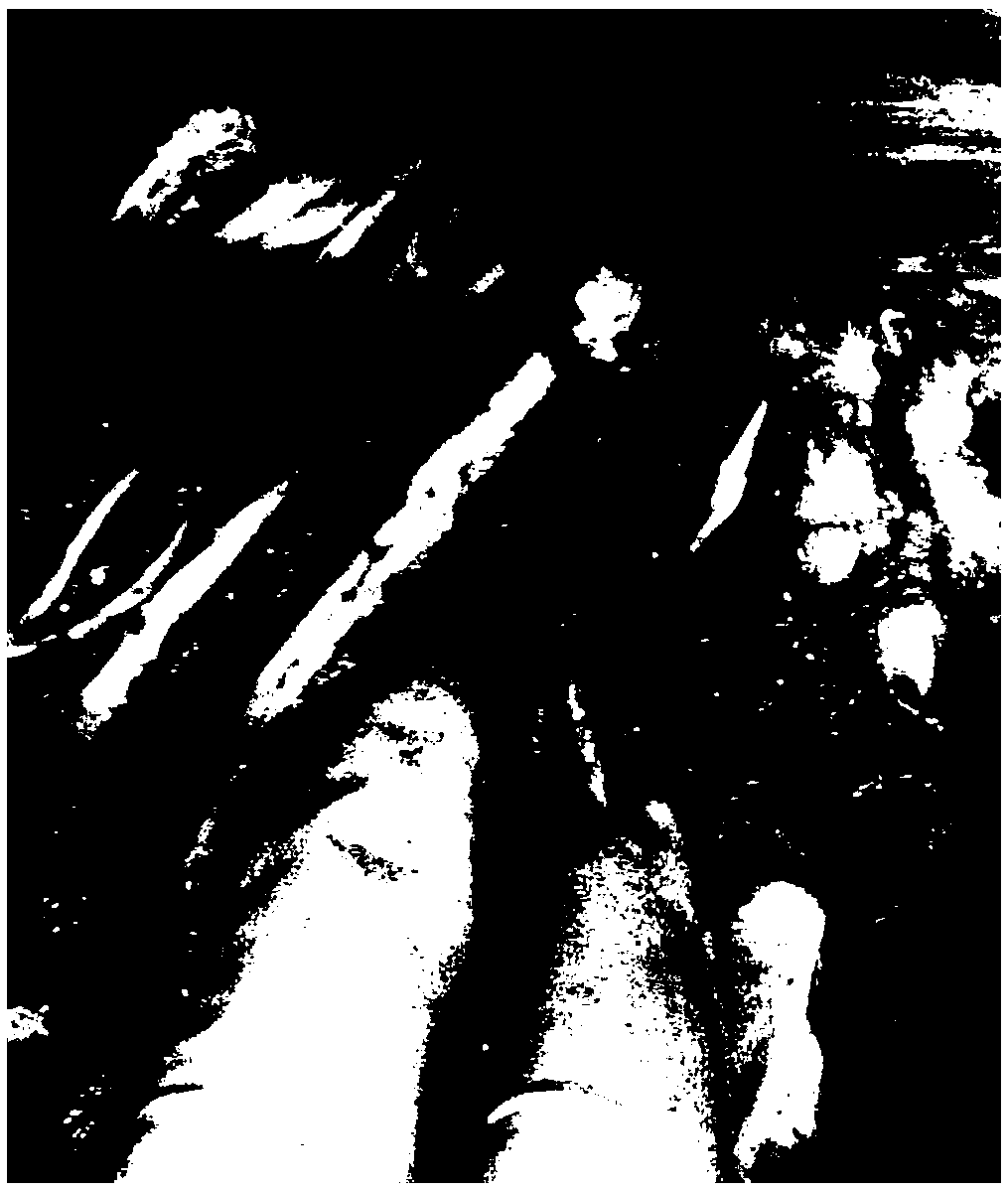
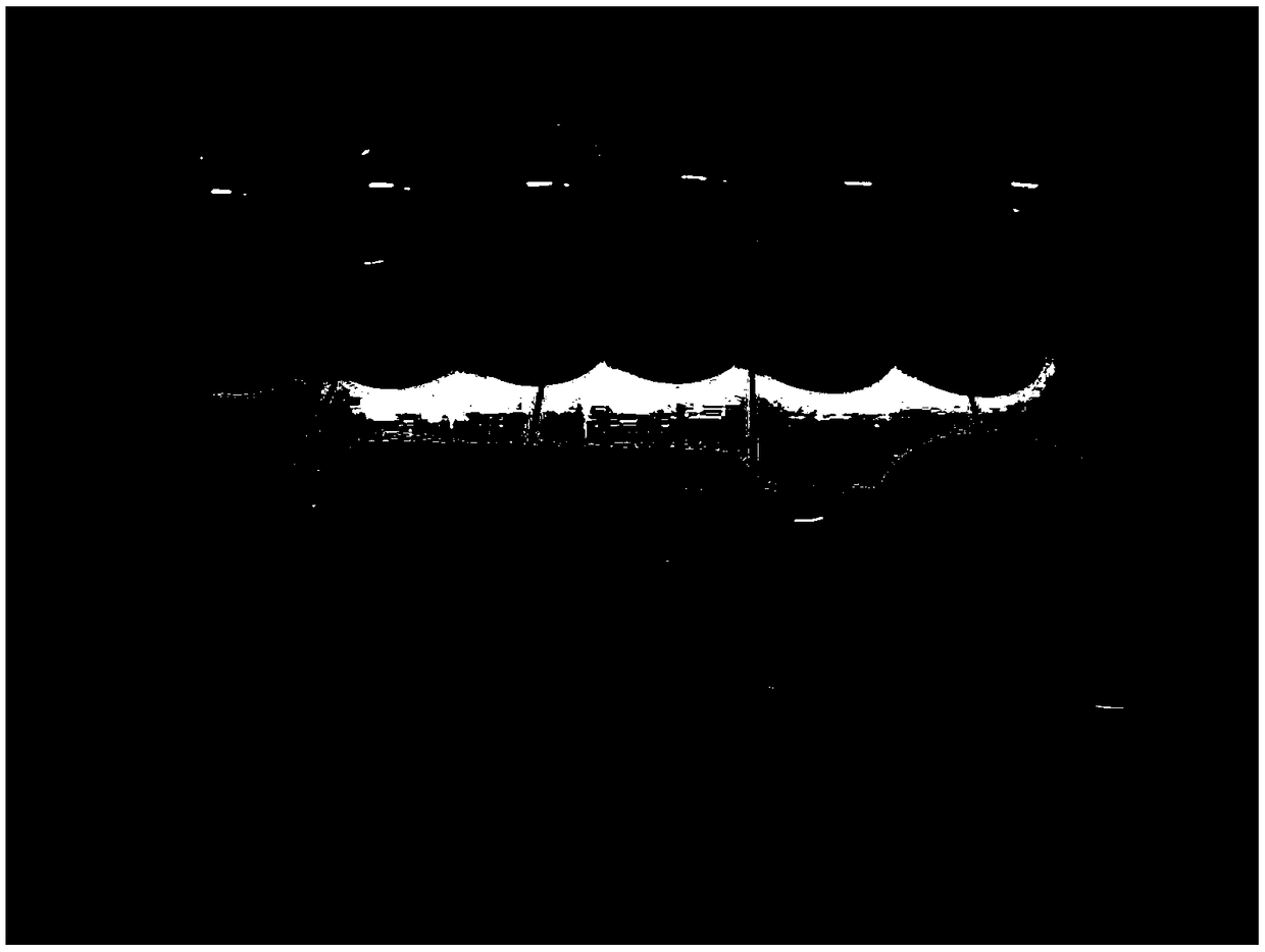
[0075] Referring to the scheme of Example 1, sub-continuous large slices of the whole mammary gland were made. Taking photos during production Figure 1-8 Shown, respectively, in the process of making sub-continuous large slices of the whole mammary gland figure 1 draw materials, figure 2 location markers, image 3 Gradient ethanol dehydration, Figure 4 xylene transparent, Figure 5 Dip Wax, Figure 6 Wax-soaked tissues, Figure 7 Embedded tissue, Figure 8 Tissue sections after HE staining.

[0076] The specific production process is as follows: collect tumor specimens from patients with invasive ductal carcinoma of the breast, and after complete mastectomy (before dissection of axillary lymph nodes), the surgeon will completely enucleate the tumor from the breast specimen, and cut off the surrounding obvious excess Adipose and normal glandular tissue. A small piece about 5×5×2.5mm (length×width×height) in the middle of the tumor was taken and sent to the patholog...

Embodiment 3

[0081] Summary Example 1-2 utilizes small-interval continuous histopathological sections of whole breast tumors to compare with conventional ones. There is no significant difference in HE staining results during the preparation process of Example 1-2, but it solves the problem of making large breast pathological sections. A series of technical problems occurred during the process, such as poor fixation effect, bending deformation, thick slices, poor quality, easy staining, and inability to observe under ordinary upright microscopes. The main reason is that during the production process, corresponding pretreatment preparations are carried out for the whole breast tumor specimen, which makes the sample dehydration, transparency, wax immersion and other effects better, and can achieve a better comprehensive treatment effect of the sample, making subsequent large slice production easier and more efficient. .

[0082] It also effectively shortens the pre-preparation time from 7 day...


PUM


Abstract

The invention discloses a whole breast subserial large slice preparation method. The method comprises the following steps of collecting a tumor sample; performing continuous parallel splitting to obtain sample blocks which are split in parallel; performing soaking and fixing in a neutral formaldehyde solution with the concentration of 4% for 24 hours; removing redundant fat tissues by clipping; performing continuous parallel cutting in parallel to an original slice again to form a slice shape; performing continuous fixing for 48 hours by using the neutral formaldehyde solution with the concentration of 4%; performing sequential dehydration by adopting gradient ethanol; performing transparency treatment by using xylene; performing wax dipping in an electric heating constant temperature incubator at 62-65 DEG C to obtain a paraffin pathological sample; and slicing the paraffin pathological sample by adopting a rotary paraffin slicing machine, wherein each sample block can be subjected toconventional pathological staining including HE through continuous slicing. The thickness of a large slice prepared with the method can reach the conventional slice thickness of 4-5 microns; the slice is complete in tissue structure, smooth, clean and free of folds and pollution; no slice stripping phenomenon occurs; the cell nucleoplasm proportion is clear; and a staining result is consistent with a conventional clinical staining result.

Description

technical field [0001] The invention relates to a method for making pathological slices, in particular to a method for making continuous pathological slices at small intervals of whole breast tumors, which belongs to the technical field of medical teaching and clinical medical experiment research, in particular to scientific research on pathological experiment technology research. Background technique [0002] Breast pathological large section refers to the unconventional pathological slide production technology that cuts the whole breast and tumor specimens of breast cancer patients surgically removed in a certain thickness and evenly as a whole, collects materials, and finally makes large pathological sections. The slices prepared by this technology can observe the whole picture of the tumor and its surrounding breast tissue, and can also observe the growth characteristic change and expansion mode of the whole tumor in the breast from different sections. It can not only me...

Claims


Application Information

Patent Timeline
Patent Type & Authority Applications(China)
IPC IPC(8): G01N1/06G01N1/30
CPCG01N1/06G01N1/30
Inventor 陈洁王竹王晓璐黄芹王艳萍
Owner WEST CHINA HOSPITAL SICHUAN UNIV
Why Eureka
Try Eureka