Method for detecting rare cells in blood based on surface enhancement effect

A rare cell, surface-enhanced technology, applied in the field of medical diagnosis, can solve the problems of false negatives and low capture efficiency of CTC cells, and achieve the effect of high capture efficiency, improved capture ability, and enhanced detection signal.

Inactive Publication Date: 2018-12-21
NINGBO MEIJING MEDICAL TECH
View PDF4 Cites 3 Cited by
  • Summary
  • Abstract
  • Description
  • Claims
  • Application Information

AI Technical Summary

Problems solved by technology

[0005] Among them, in the selection of CTC chips, from the original selection of ordinary glass slides to the selection of aldehyde-modified glass slides or epoxy resin-coated glass slides,

Method used


Image

  • Method for detecting rare cells in blood based on surface enhancement effect
  • Method for detecting rare cells in blood based on surface enhancement effect
  • Method for detecting rare cells in blood based on surface enhancement effect

Examples


Embodiment 1

[0033] A method for detecting rare cells in blood based on the surface enhancement effect of the present invention comprises the following steps:

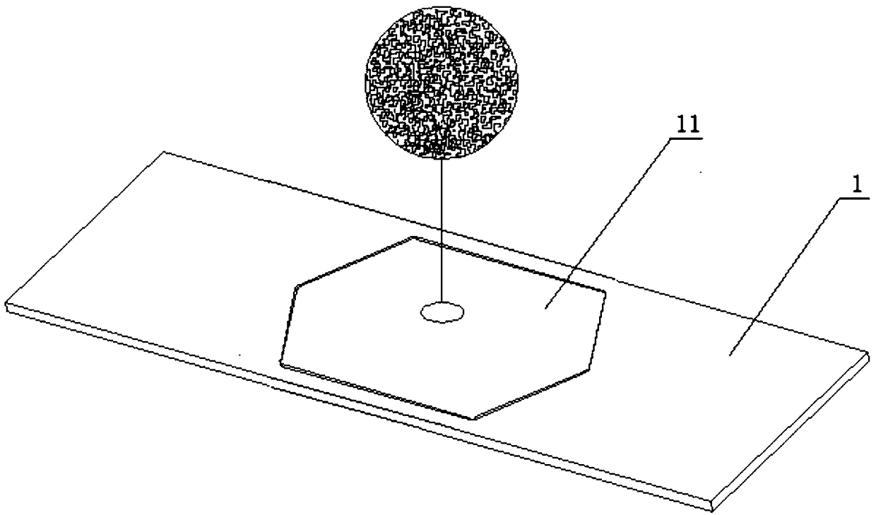
[0034] (1) Coating a layer of plasma non-ferrous metal film 11 on the surface of the glass slide by spraying to prepare a metal nanostructured SERS (Surface-enhanced Raman scattering, surface-enhanced Raman scattering) chip 1;

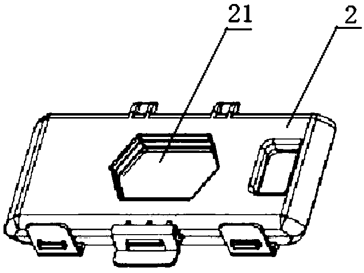
[0035] (2) Install the metal nanostructured SERS chip 1 into the capture box 2 of the capture instrument 3, and install the matching capture consumables package and supporting reagents;

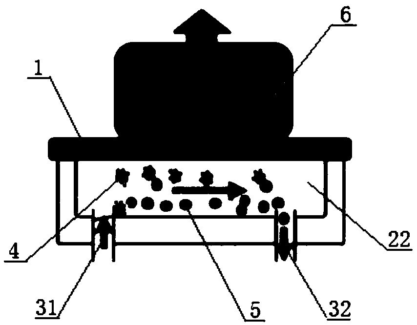
[0036] (3) Load the pretreated blood sample into the capture instrument 3, set the operating parameters of the capture instrument 3, and use the immunomagnetic enrichment method to capture rare cells in the blood sample;

[0037] (4) taking the captured metal nanostructure SERS chip 1 out of the capture box 2, drying at 37°C, and performing immunofluorescence staining on the metal nanostructure SERS chip 1 after drying;

...

Embodiment 2

[0043] Taking the tumor cell MCF-7 simulated sample as an example, an ordinary chip (that is, a glass slide without a plasma non-ferrous metal film 11 on the surface) is used as a control group, and a gold-plated SERS chip of the present invention (wherein the SERS chip 1 of the metal nanostructure adopts The metal material is gold) as the experimental group, and the MCF-7 cells were captured and detected by fluorescence.

[0044] (1) MCF-7 cells were cultured, collected and diluted for later use.

[0045] (2) The blood of a healthy person is taken, and MCF-7 is added to it to prepare a simulated sample.

[0046] (3) The simulated sample is equally divided into two samples to be tested: the sample to be tested in the experimental group and the sample to be tested in the control group, each sample to be tested is 5ml.

[0047] (4) The immune magnetic particle capture instrument 3 is powered on and self-inspected, and the ordinary chip and the gold-plated SERS chip are respecti...

Embodiment 3

[0055] Taking the peripheral blood samples of 13 tumor patients as an example, ordinary chips, cadmium-coated SERS chips and gold-coated SERS chips were used for CTC cell capture and fluorescence detection.

[0056] (1) Take the peripheral blood of 13 tumor patients in ACD blood collection tubes, subpackage and test each sample within 24 hours, 5ml / tube, divided into 3 tubes.

[0057] (2) After the blood sample is pretreated by the sample pretreatment solution, immunomagnetic particles are added and loaded into the capture device 3 .

[0058] (3) Set the operating parameters of the capture instrument 3: the magnetic particle capture rate is 2.5ml / hr, the chip cleaning rate is 5ml / hr, and the instrument is operated.

[0059] (4) After capturing the CTC cells, remove the capture box 2 from the instrument, open it, take out the ordinary chip, the cadmium-plated SERS chip and the gold-plated SERS chip, and dry it at 37°C.

[0060] (5) The dried cadmium-plated SERS chip and the go...


PUM

PropertyMeasurementUnit
Thicknessaaaaaaaaaa
Thicknessaaaaaaaaaa

Abstract

The invention discloses a method for detecting rare cells in blood based on surface enhancement effect, which is characterized by comprising the steps of: plating a layer of plasma colored metal membrane on the surface of slide using spray coating method, and obtaining a SERS chip with metal nanostructure; arranging the SERS chip into a capture box of a capture apparatus; loading a pretreated blood sample in the capture apparatus, setting operating parameters of the capture apparatus, and capturing rare cells in the blood sample by an immune-magnetic enrichment method; taking the SERS chip with metal nanostructure out of the capture box after capturing, and performing immunofluorescence staining on the SERS chip after drying at 37 DEG C; and scanning and detecting the stained rare cells using a detection system. According to the method for detecting rare cells in blood based on surface enhancement effect, the capturing efficiency of rare cells such as CTC cells in the blood can be obviously improved, and the detection signal of rare cells can be enhanced

Description

technical field [0001] The invention relates to the field of medical diagnosis, in particular to a method for detecting rare cells in blood based on a surface enhancement effect. Background technique [0002] Malignant tumors seriously threaten human health, and the main cause of death is that some tumor cells acquire the ability to invade and cause distant metastasis. Tumor metastasis is a complex process involving multi-step factors. Tumor cells with metastatic potential break away from the primary tumor and metastasize to the blood, bone marrow, lymph nodes or distant organs in very small numbers, which are difficult to detect using conventional detection methods. for tumor micrometastases. The shedding, invasion and entry of tumor cells into the blood circulation are the initial stages of tumor metastasis, and provide the possibility for the formation of tumor metastases. Therefore, the detection of circulating tumor cells (CTC) in peripheral blood has important clinica...

Claims


Application Information

Patent Timeline
IPC IPC(8): G01N21/65G01N33/533G01N33/543
CPCG01N21/658G01N33/533G01N33/5436
Inventor 张晓晶沈挺
Owner NINGBO MEIJING MEDICAL TECH
Why Eureka
Try Eureka