Lupus anticoagulant detection reagent as well as preparation method and use method thereof

A lupus anticoagulant and detection reagent technology, applied in the field of biological detection reagents, can solve the problems of increased false positive rate and false negative rate of test results, no LA, poor stability, etc., to reduce false positive rate and false negative rate, The effect of improving accuracy and low cost

Inactive Publication Date: 2019-03-26
THE FIRST AFFILIATED HOSPITAL OF ARMY MEDICAL UNIV
View PDF1 Cites 6 Cited by
  • Summary
  • Abstract
  • Description
  • Claims
  • Application Information

AI Technical Summary

Problems solved by technology

[0006] (1) At present, there is no reference material and reference method for LA detection, which poses a great challenge to experimental test personnel to ensure the accuracy of test results
[0007] (2) Sin

Method used


Image

  • Lupus anticoagulant detection reagent as well as preparation method and use method thereof
  • Lupus anticoagulant detection reagent as well as preparation method and use method thereof
  • Lupus anticoagulant detection reagent as well as preparation method and use method thereof

Examples


preparation example Construction

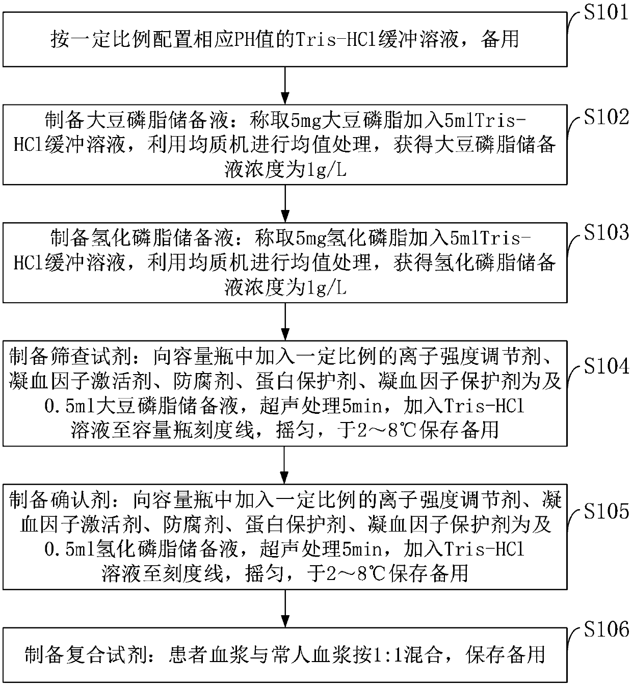
[0045] Such as figure 1 As shown, a preparation method for detecting lupus anticoagulant reagent provided by the embodiment of the present invention specifically includes the following steps:

[0046] S101: configure the Tris-HCl buffer solution of the corresponding pH value in a certain proportion, for subsequent use;

[0047] S102: Prepare soybean lecithin stock solution: Weigh 5 mg soybean lecithin and add 5 ml Tris-HCl buffer solution, use a homogenizer to perform mean value processing, and obtain a concentration of soybean lecithin stock solution of 1 g / L;

[0048] S103: Prepare hydrogenated phospholipid stock solution: Weigh 5 mg of hydrogenated phospholipid and add 5 ml of Tris-HCl buffer solution, use a homogenizer to perform mean value processing, and obtain a hydrogenated phospholipid stock solution with a concentration of 1 g / L;

[0049] S104: Preparation of screening reagents: add a certain proportion of ionic strength regulator, coagulation factor activator, pres...

Embodiment 1

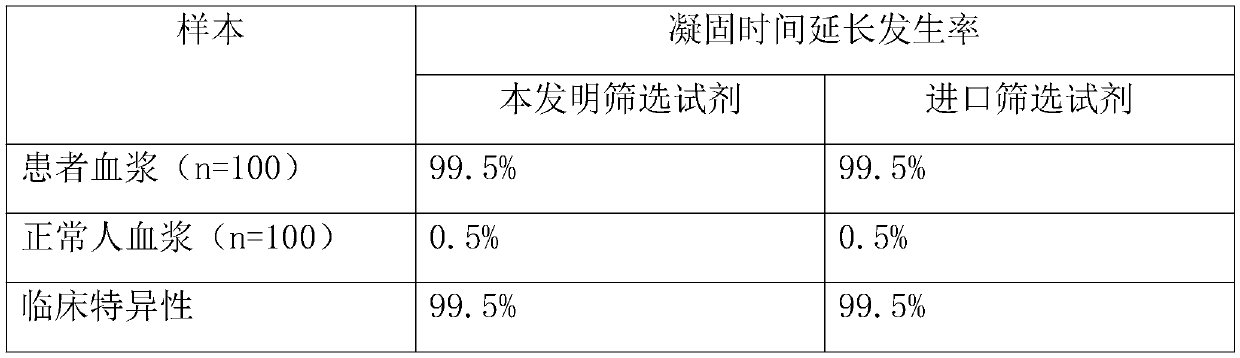
[0061] With the test kit prepared by the present invention and imported other brands of LA detection reagents, 200 cases of clinical plasma samples were respectively screened and measured for LA, and the incidence of prolongation of plasma coagulation time was counted to investigate the clinical specificity of the reagents. The results are shown in Table 1. .

[0062] Table 1

[0063]

[0064] As can be seen from Table 1, the clinical specificity of the kit of the present invention reaches the standard of imported reagents.

Embodiment 2

[0066] The kit prepared by the present invention was stored at 6°C, taken out when used, and the coagulation time of the same LA normal plasma and LA abnormal plasma was measured at different times, and the results are shown in Table 2.

[0067] Table 2

[0068]

[0069]

[0070] As shown in Table 2, the kit of the present invention has been continuously measured for 15 days, and the results have remained stable, indicating that the kit of the present invention has better stability.


PUM


Abstract

The invention belongs to the technical field of biological detection, and discloses a lupus anticoagulant detection reagent as well as a preparation method and a use method thereof. The lupus anticoagulant detection reagent mainly comprises a screening reagent, a confirming reagent and a compounding region, wherein the screening reagent is prepared from blood coagulation factor activators with thevolume percentage being 13mg/L, 65mmol/L of buffer solution with the pH being 7.5, 55mg/L of synthetic phospholipids, 75g/L of protein protection agents, 0.06 percent of preservatives, 100mmol/L of ion intensity regulators and 8g/L of blood coagulation factor protection agents; the conforming agent has high content of synthetic phospholipids. The reagent is a liquid reagent; the preparation process is simple and convenient; the cost is relatively low; through the addition of the protein protection agents, the stability of the liquid reagent is effectively ensured; the false positive rate andfalse negative rate in the detection result are effectively reduced; different synthetic phospholipids are respectively used on screening reagents and conforming reagents, so that the LA detection sensitivity is improved; the detection test accuracy is improved.

Description

technical field [0001] The invention belongs to the technical field of biological detection reagents, in particular to a lupus anticoagulant detection reagent, a preparation method and a use method thereof. Background technique [0002] At present, the existing technologies commonly used in the industry are as follows: [0003] Lupus anticoagulants (Lupus anticoagulants, LA) is a phospholipid-dependent pathological circulating anticoagulant substance, which is a heterogeneous immunoglobulin of IgG or IgM, and is complexed by combining 2-glycoprotein I, human prothrombin and other phospholipids Drugs to interfere with the phospholipid-dependent coagulation process and prolong the coagulation time. Persistent LA-positive patients are considered to have a higher risk of thrombosis and recurrence, and are also a danger signal for unexplained recurrent miscarriage, stillbirth, thrombophilia, and certain autoimmune diseases. The detection of LA includes screening test, confirmat...

Claims


Application Information

Patent Timeline
IPC IPC(8): G01N33/68
CPCG01N33/6854
Inventor 牟方祥吴红游苘霞
Owner THE FIRST AFFILIATED HOSPITAL OF ARMY MEDICAL UNIV
Why Eureka
Try Eureka