

Application of miR-18a-5p agonist in preparing anti-retinal neovascularization drugs
A technology of mir-18a-5p, 1.mir-18a-5p, which is applied in the field of preparation of anti-retinal neovascularization drugs, and can solve the problems that the effect has not been reported yet
- Summary
- Abstract
- Description
- Claims
- Application Information
AI Technical Summary
Problems solved by technology
Method used
Image

Examples
Embodiment 1
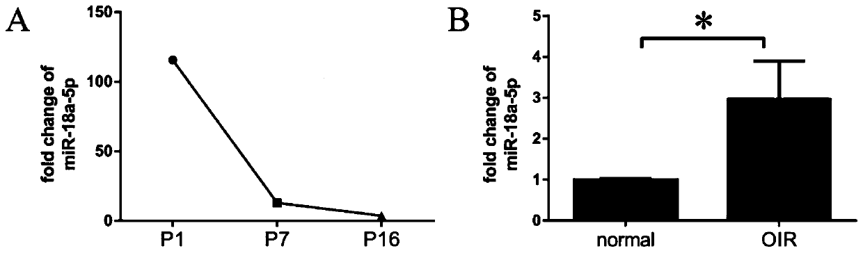
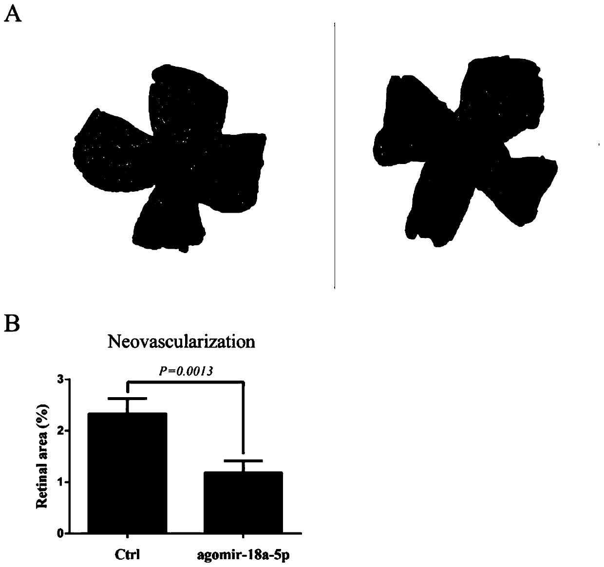
[0030] Example 1 Detection of miR-18a-5p expression level in mouse developmental stages and OIR model
[0031] Firstly, wild-type C57BL / 6J mice were used to establish the OIR model, which is a classic animal model for the study of retinal neovascularization. Use Omega's miRNA Kit kit to extract total RNA from the retina of the developmental stage (embryo (E) 14.5 days, postnatal (P) 1, 7, 16 days) and OIR model mice. The main steps are as follows: first, the extracted retina Place in a 1.5ml centrifuge tube, add 1ml RNA-solve Reagent, place at room temperature for 3 minutes, and use a tissue homogenizer to lyse; then add 0.2ml chloroform, mix vigorously for 15 seconds, place on ice for 10 minutes; then centrifuge at 4°C, 12000g for 15 minutes minutes, transfer no more than 80% of the total volume of the colorless upper liquid to a new 1.5ml centrifuge tube; add 1.5 times the volume of absolute ethanol, mix thoroughly and add to the filter column (provided in the kit), centrifu...
Embodiment 2
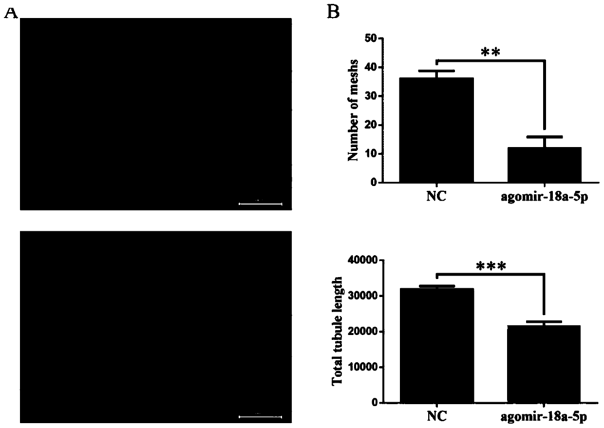
[0034] Example 2 Effect of miR-18a-5p on the formation of blood vessels by HRMEC
[0035] Firstly, cells were transfected with miR-18a-5p agonist, which was provided by Ribo. Plates were coated with Bovine Plasma Fibronectin (BPF) prior to seeding cells. HRMECs were seeded in 24-well plates 24 hours before transfection, and transfection was performed when adherent cells covered 60% of the bottom area. The culture medium used is Endothelial Cell Medium (ECM), supplemented with 5% calf serum, 20ng / ml Endothelial Cell Growth Supplement (ECGS) and 1% P / S. Aspirate the original cell culture medium before transfection, add fresh serum-free culture medium, and place in an incubator (37°C, 5% CO 2 ) for half an hour. Dissolve miR-18a-5p agonist (40nM) in serum-free culture medium, and mix Lipofectamine RNAiMAX Reagent in an equal volume of serum-free culture medium at the same time, mix the two liquids, and let stand for 15 minutes. Slowly add this transfection solution to the cel...
Embodiment 3
[0037] Example 3 miR-18a-5p inhibits the expression of FGF1
[0038] The agomir-18a-5p was transfected into HUVEC, and the total RNA was extracted as in Example 2. Take 0.5 μg total RNA, add 1 μl Random Primer and 1 μl Oligo(dT). Mix well, put into a PCR instrument at 70°C for 5 minutes, and place on ice after the reaction. Configure the reaction system, take 6.1μl DEPC water, GoScript 5x Reaction Buffer 4μl, MgCl 2 2.4 μl, PCR Nucleotide Mix 1 μl, Recombinant RNasin Ribonuclease Inhibitor 0.5 μl, and GoScript Reverse Transcriptase 1 μl. Take 15 μl of reaction system and 5 μl of total RNA, mix thoroughly, and put them into a PCR instrument for amplification. The reaction conditions are: 25°C for 5 minutes; 42°C for 1 hour; 70°C for 15 minutes. Take 100 ng of cDNA, 5 μl of iTaqTM Universal SYBR Green Supermix buffer, 1.5 μl of Forward and Reverse primer, and make up the volume to 10 μl with DEPC water. After mixing well, place in QuantStudio TM 5Real-Time PCR Systems, run...
PUM
 Abstract
Description
Claims
Application Information