Leukemia marker joint detection antibody chip and kit

An antibody chip and leukemia technology, applied in the field of biomedicine, can solve problems such as difficulties, achieve reliable results, improve accurate diagnosis, and use less specimens

Active Publication Date: 2022-05-17
RAYBIOTECH INC GUANGZHOU +1
View PDF5 Cites 0 Cited by
  • Summary
  • Abstract
  • Description
  • Claims
  • Application Information

AI Technical Summary

Problems solved by technology

[0004] Since the regulation of cell signaling pathways is a network structure, and because cytokines form a network with each other in the body and interact with various immune cells to participate in the development of leukemia, it is necessary to simultaneously measure the changes in the content of multiple cytokines in order to respond comprehensively and accurately The mechanism of leukemia occurrence and progression, while traditional ELISA and flow cytometry techniques can only detect certain factors, and cannot understand the change level of leukemia biomarkers as a whole and comprehensively, in-depth understanding of leukemia pathogenesis, and for leukemia It is very difficult in terms of accurate diagnosis, disease course monitoring and prognosis assessment

Method used


Image

  • Leukemia marker joint detection antibody chip and kit
  • Leukemia marker joint detection antibody chip and kit
  • Leukemia marker joint detection antibody chip and kit

Examples


Embodiment 1

[0031] Example 1 Preparation of the Antibody Chip Kit for the Joint Detection of Leukemia Markers

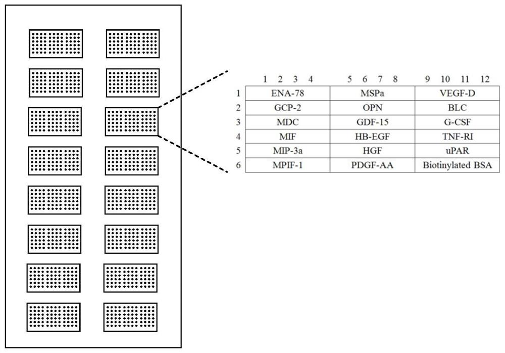
[0032] 1. Leukemia markers and their specific antibodies:

[0033] Preparations immobilized with recombinant human epithelial granulocyte activation protein 78 (ENA-78), neutrophil chemoattractant protein-2 (GCP-2,), macrophage-derived chemokine (MDC), macrophage migration inhibition factor (MIF), macrophage inflammatory protein-3a (MIP-3a), myeloid progenitor inhibitory factor-1 (MPIF-1), macrophage stimulating protein a (MSPa), osteopontin (OPN), growth Differentiation factor 15 (GDF-15), heparin-binding epidermal growth factor (HB-EGF), human growth factor (HGF), recombinant human platelet-derived growth factor-AA (PDGF-AA), vascular endothelial growth factor D (VEGF- D), B lymphocyte chemotactic factor (BLC), granulocyte colony-stimulating factor (G-CSF), tumor necrosis factor R1 (TNF-RI), urokinase-type plasminogen activator receptor (uPAR) leukemia The chip of the specif...

Embodiment 2

[0060] Example 2 Leukemia marker joint detection antibody chip kit

[0061] In addition to the antibodies described in Example 1, the leukemia marker joint detection antibody chip in the leukemia marker joint detection antibody chip kit described in this example is also immobilized with anti-tumor necrosis factor (TNFa), fas receptor (Fas L), interleukin 4 (IL-4), intercellular adhesion molecule 1 (ICAM-1), interleukin 5 (IL-5), growth-regulated oncogene 1 (KC), inhibitor of matrix metalloproteinase 1 (TIMP-1), human thymus activation Regulated chemokine (TARC), interleukin 2 (IL-2), interleukin 7 (IL-7), macrophage colony-stimulating factor (M-CSF), interleukin 10 (IL-10), interleukin 21 (IL-21 ), interleukin 12 (IL-12p70), brain-derived neurotrophic factor (BDNF), platelet factor 4 (PF-4), epidermal growth factor (EGF), tumor necrosis receptor II (TNF-RII), tumor necrosis factor A total of 36 specific antibodies for leukemia biomarkers such as superfamily 14 (LIGHT); corres...

Embodiment 4

[0062] Example 4 Experiment of Quantitative Detection of Leukemia Markers

[0063] 1. Complete drying of the slide antibody chip

[0064] Take the slide antibody chip described in Example 1 out of the box, and after equilibrating at room temperature for 20-30 minutes, open the packaging bag, peel off the sealing strip, and then place the chip in a vacuum desiccator or dry it at room temperature for 1-2 hours .

[0065] 2. Gradient dilution of the leukemia marker standard mixture:

[0066] 2.1. Add 500 μl of the sample diluent described in Example 1 to the vial of the leukemia marker standard mixture, and redissolve the standard. Before opening the vial, give it a quick centrifuge and gently pipet up and down to dissolve the powder. Label this vial as Std 1.

[0067] 2.2. Mark 6 clean centrifuge tubes as Std2, Std3 to Std7 respectively, and add 200 μl of sample diluent to each small tube.

[0068] 2.3. Take 100 μl of Std 1 and add it to Std2 and mix gently, then take 100 μl...


PUM


Abstract

The invention relates to an antibody chip for joint detection of leukemia markers, wherein the leukemia markers are selected from: ENA-78, GCP-2, MDC, MIF, MIP-3a, MPIF-1, MSPa, OPN, GDF-15, HB-EGF , HGF, PDGF‑AA, VEGF‑D, BLC, G‑CSF, TNF‑RⅠ, uPAR, TNFa, Fas L, IL4, ICAM‑1, IL5, KC, TIMP‑1, TARC, IL2, IL7, M‑ At least 17 of CSF, IL10, IL21, IL‑12p70, BDNF, PF‑4, EGF, TNFRⅡ, and LIGHT can simultaneously measure the expression levels of multiple samples and multiple leukemia markers to accurately and comprehensively reflect the occurrence and course of leukemia , to improve its diagnosis, disease course monitoring and prognosis assessment.

Description

technical field [0001] The invention belongs to the technical field of biomedicine, in particular to an antibody chip for quantitatively detecting leukemia markers simultaneously. Background technique [0002] Leukemia, also known as blood cancer, is a malignant tumor of the hematopoietic system. The source of the disease is the abnormal work of the hematopoietic tissue in the bone marrow due to the mutation of DNA in the cells. Stem cells in the bone marrow make thousands of red and white blood cells each day. Leukemia patients overproduce immature white blood cells, hindering other work of the bone marrow, which reduces the function of the bone marrow to produce other blood cells. Leukemia can spread to the lymph nodes, spleen, liver, central nervous system, and other organs. [0003] Leukemia is a kind of malignant clonal disease of hematopoietic stem cells. The incidence of leukemia in various regions of my country ranks sixth among all kinds of tumors. Acute myeloid...

Claims


Application Information

Patent Timeline
Patent Type & Authority Patents(China)
IPC IPC(8): G01N33/574
Inventor 张惠华吕跃朱思为匡治州黄若磐
Owner RAYBIOTECH INC GUANGZHOU
Why Eureka
Try Eureka