Trichophyton rubrum infection model preparation method

A technology of Trichophyton rubrum and models, applied in biochemical equipment and methods, measurement/testing of microorganisms, epidermal cells/skin cells, etc., can solve the problems that are not close to the biological state of skin fungal infection, and animal models are not easy to observe, etc.

Pending Publication Date: 2019-07-05
HOSPITAL OF DERMATOLOGY CHINESE ACAD OF MEDICAL SCI
View PDF5 Cites 4 Cited by
  • Summary
  • Abstract
  • Description
  • Claims
  • Application Information

AI Technical Summary

Problems solved by technology

[0004] For this reason, the embodiment of the present invention provides a preparation method of a Trichophyton rubrum infection model to solve the problem th

Method used


Image

  • Trichophyton rubrum infection model preparation method
  • Trichophyton rubrum infection model preparation method
  • Trichophyton rubrum infection model preparation method

Examples


Embodiment 1

[0023] Embodiment 1 The preparation method of the Trichophyton rubrum infection model of the embodiment of the present invention

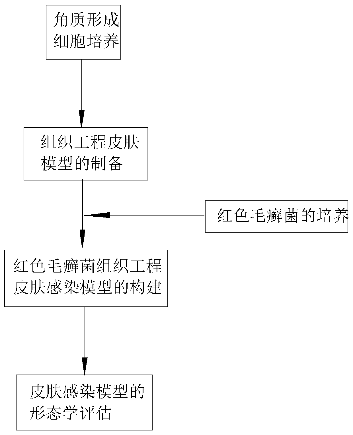
[0024] Such as figure 1 Shown, in the implementation of the present invention, the preparation method of Trichophyton rubrum infection model comprises the following steps:

[0025] 1. Acquisition and in vitro culture of human keratinocytes

[0026] Take healthy foreskin tissue, firstly soak and disinfect the skin tissue with iodophor for 1-3 minutes in the aseptic operation table, then deiodine and disinfect with 75% alcohol for 2 minutes, and finally rinse with PBS containing 1% cyano-chain double antibody for 3 times;

[0027] Use scissors and a scalpel blade to remove the subcutaneous fat layer and connective tissue of the foreskin as much as possible, and cut the skin into 2cm 2 Digested overnight at 4°C in 10mg / ml Dispase II;

[0028] The next day, the digested skin was separated from the dermis and epidermis with tweezers, and the separate...

Embodiment 2

[0034] Embodiment 2 Utilize the tissue engineering skin model of the embodiment of the present invention to establish the Trichophyton rubrum infection model

[0035] 1. Construction of Trichophyton rubrum tissue-engineered skin infection model

[0036] The standard strain of Trichophyton rubrum, ATCCMYA4438, was purchased from the American type culture collection (Americantype culture collection), and was preserved by the Institute of Skin Research (Medical Fungi Collection) of the Pathogenic Microbial (Virus) Species Preservation Center, Chinese Academy of Medical Sciences, in SDA Cultured on the culture medium at 28°C for 15 days. Before inoculation, add an appropriate amount of sterile normal saline to the surface of the bacteria, gently scrape the surface of the bacteria with a disposable coating stick, avoid hanging on the agar with force, absorb the bacteria liquid, filter to remove the mycelium, count, and adjust the concentration to 2 ×10 5 / ml for use.

[0037] Ta...


PUM


Abstract

An embodiment of the invention discloses a trichophyton rubrum infection model preparation method and application. The preparation method includes the steps: separating a corium layer and a cuticularlayer of prepuce tissues, and digesting and culturing the cuticular layer to obtain human keratinocytes; finally, adding the human keratinocytes to a collagen scaffold and culturing the human keratinocytes to form a tissue engineering skin model. The model is simple to operate and easy to observe and solves the problem that a current trichophyton rubrum infection animal model is not easily observed. Seeds of the trichophyton rubrum infection model are the human keratinocytes and closer to the biological state of human skin fungal infection.

Description

technical field [0001] The embodiment of the present invention relates to the technical field of cell and tissue engineering, in particular to a method for preparing a Trichophyton rubrum infection model. Background technique [0002] At present, Trichophyton rubrum is the most common human-friendly dermatophyte in clinical practice, because the fungus has the characteristics of being friendly to humans. In the process of studying the mechanism of Trichophyton rubrum infecting animals, an animal infection model was chosen for research, but The construction process of animal models is complicated and difficult, and it is far from clinical infection conditions. [0003] In recent years, with the continuous in-depth research and rapid development of tissue-engineered skin, it is more convenient than other models to simulate and construct tissue-engineered skin infection models in vitro to study the pathogenesis of Trichophyton rubrum infection, the immunopathological process af...

Claims


Application Information

Patent Timeline
IPC IPC(8): C12N5/071C12Q1/02
CPCC12N5/0625C12N2533/54G01N33/5005
Inventor 刘维达王琼郑海林梅嬛彭靖雯
Owner HOSPITAL OF DERMATOLOGY CHINESE ACAD OF MEDICAL SCI
Why Eureka
Try Eureka