Method and application for promoting cells to secrete exosomes

A cell secretion and exosome technology, applied in the field of promoting cell secretion of exosomes, can solve problems such as single function and limited ability to secrete exosomes, achieve high yield, promote tissue repair function, and promote rapid wound repair. Effect

Active Publication Date: 2019-08-16
SHANGHAI JIAO TONG UNIV
View PDF3 Cites 10 Cited by
  • Summary
  • Abstract
  • Description
  • Claims
  • Application Information

AI Technical Summary

Problems solved by technology

However, the ability of cells to secrete exosomes under conventional culture conditions is limited, and their functions are often single

Method used


Image

  • Method and application for promoting cells to secrete exosomes
  • Method and application for promoting cells to secrete exosomes
  • Method and application for promoting cells to secrete exosomes

Examples


Embodiment 1

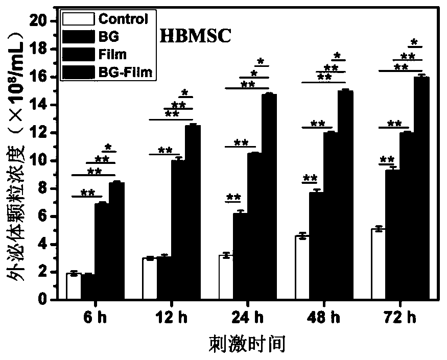
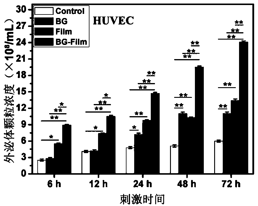
[0041] Exosomes produced by cells stimulated with biomaterial activity signals:

[0042] Step 1, prepare bioactive glass (BG) ion extraction solution, respectively add 1g BG powder to 5mL serum-free mesenchymal stem cell culture medium (MSCM) and 5mL serum-free endothelial cell culture medium (ECM), in the cell Incubate in an incubator for 24 hours, collect the supernatant, and filter the filtrate through a 0.22 μm sterile filter to obtain a BG ion extract, which is stored at 4°C for use.

[0043] Step 2, preparing directional electrospinning fiber membrane, dissolving PCL and PDLLA with a mass ratio of 1:1 in DMF and THF with a volume ratio of 4:1 at a mass volume ratio of 4.8%, and using an electrospinning device to 10kv Electrospinning was carried out under the conditions of voltage, 0.02mL / min flow rate and 12cm receiving distance, and the oriented electrospun fiber membrane was received by a high-speed drum rotating at 2000r / min, which was cut into discs with a diameter o...

Embodiment 2

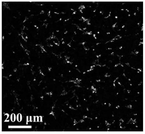
[0052] Verification of the ability of HBMSC-derived exosomes to promote HUVEC angiogenic differentiation in vitro:

[0053] According to the method shown in Example 1, the exosomes secreted by HBMSCs under BG stimulation for 48 hours and under normal conditions were collected, respectively recorded as HBMSC-BG-exosome and HBMSC-Control-exosome, and stored at 4°C for future use.

[0054] Digest passage HUVEC, according to 4 × 10 4 HUVEC / cm2 cell density were inoculated into sterile 6-well culture plate and 48-well plate covered with cell slides respectively, cultured with complete ECM for 24 hours, washed with PBS three times, and HBMSC-BG- exosome and HBMSC-Control-exosome diluted to 1 × 10 8 pcs / mL, add 2 mL of HBMSC-BG-exosome exosome dilution to each well of the first 6-well plate, add 2 mL of HBMSC-Control-exosome exosome dilution to each well of the second 6-well plate, No exosome dilution solution was added to the third 6-well plate, 0.5 mL of HBMSC-BG-exosome exosome ...

Embodiment 3

[0058] Verification of the ability of HBMSC-derived exosomes to promote HUVEC subcutaneous angiogenesis in vivo:

[0059] According to the method shown in Example 1, the exosomes secreted by HBMSCs under BG stimulation for 48 hours and under normal conditions were collected, respectively recorded as HBMSC-BG-exosome and HBMSC-Control-exosome, and stored at 4°C for future use.

[0060] On the back of three Kunming mice, a small area of ​​hair was shaved with a shaving machine, and the first mouse was injected with 50 μL of particles with a concentration of 1×10 8 / mL PBS solution of HBMSC-BG-exosome, the second injection of 50 μL containing particles with a concentration of 1×10 8 / mL of the PBS solution of HBMSC-Control-exosome, the third mouse was injected with PBS solution without exosomes as the control (Control), and after 1 week, the experimental mice were killed by neck dislocation, and the skin of the exosome injection area was taken, and the The inner tiny blood vesse...


PUM


Abstract

The invention discloses a method and application for promoting cells to secrete exosomes, and relates to the fields of biological materials, tissue engineering and regenerative medicines. According tothe method, the cells are stimulated by active signals of a biological material, and the exosomes generated by the cells are collected, and are used for tissue wound treatment. The operation route ofthe method and the application scheme of the obtained exosomes are described in detail, under the stimulation effects of bioactive glass chemical signals, electrostatic spinning fiber membrane structure signals and chemical/structural combination signals, more exosome particles can be secreted by mesenchymal stem cells and umbilical vein endothelial cells, and the exosomes can promote the corresponding target cells to express higher levels of functional genes, secrete more multifunctional proteins and carry out deep functional differentiation, so that the tissue wound surface can be rapidly repaired. The exosomes obtained by the method disclosed by the invention have the characteristics of higher yield and stronger tissue repair promotion function.

Description

technical field [0001] The invention relates to the fields of biomaterials, tissue engineering and regenerative medicine, in particular to a method and application for promoting exosome secretion by cells. Background technique [0002] Tissue trauma has always been faced with a large number of patients in clinical practice. Although human tissue has a certain self-repair ability, when the trauma area is too large, the patient is weak, the body function is poor, or the local environment of the wound is disordered, it is not conducive to its self-repair, such as When the blood vessels are severely damaged and the supply of blood and nutrients is insufficient, the wound cannot be repaired normally, and the wound that has not been repaired normally for a long time not only seriously affects the quality of life of the patient, but may even cause other diseases in the body, further endangering the health of the patient. Therefore, finding a method that can promote rapid and effect...

Claims


Application Information

Patent Timeline
Patent Type & Authority Applications(China)
IPC IPC(8): C12N5/0775A61L27/38A61L27/18
CPCC12N5/0663A61L27/3834A61L27/18C12N2533/30C12N2533/40C12N2533/12
Inventor 李海燕吴治
Owner SHANGHAI JIAO TONG UNIV
Why Eureka
Try Eureka