

Cell preparation for treating inflammatory enteritis
A cell preparation and cell technology, applied in cell culture active agents, animal cells, vertebrate cells, etc., can solve the problems of adverse reactions of patients, postoperative recurrence, and limited efficacy in critical cases.
- Summary
- Abstract
- Description
- Claims
- Application Information
AI Technical Summary
Problems solved by technology
Method used
Image

Examples
Embodiment 1
[0111] Example 1 Obtaining Umbilical Cord Wharton Section Mesenchymal Stem Cells
[0112] 1. Fetal umbilical cord acquisition
[0113] The umbilical cords of full-term newborns without congenital diseases were taken; the mothers had no infectious diseases such as hepatitis, syphilis, and AIDS, and the mothers and their families gave informed consent to the use of umbilical cords in the experimental research.
[0114] 2. Obtaining mesenchymal cells from the Wharton section of the umbilical cord
[0115] In a sterile laboratory bench, the umbilical cord was repeatedly washed with phosphate buffered saline (PBS) to remove residual blood. Cut the umbilical cord into small sections of 2-3cm with sterile surgical instruments, cut the umbilical cord longitudinally, remove the umbilical artery, umbilical vein and amniotic membrane, and cut the Wharton area into 0.5-1mm 3 Little pieces left and right. Obtain primary umbilical cord mesenchymal cells by tissue block culture method: sp...
Embodiment 2
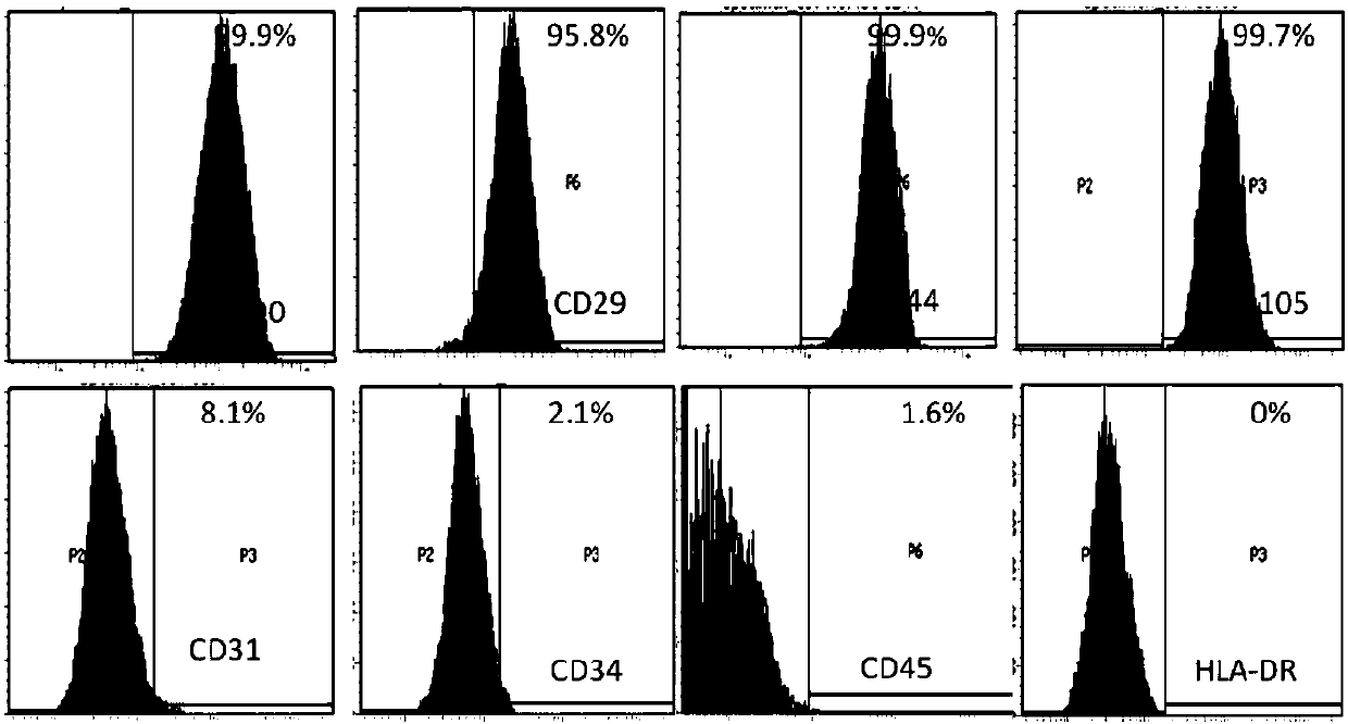
[0116] Example 2 Obtaining Umbilical Cord Wharton Section Mesenchymal Stem Cell Preparation
[0117] The above-mentioned low-serum medium was used to culture Example 1 to obtain umbilical cord Wharton interval mesenchymal stem cells. Specifically, the human umbilical cord mesenchymal stem cells isolated by the tissue block method were cultured in the above-mentioned low serum medium, and when the cells were about 80% confluent, they were digested with 0.25% trypsin for 1-2 minutes, and most of the cells were used for expansion. In culture, a small fraction of cells is used to identify markers of stem cells. The expression of CD29, CD44, CD90, CD105, CD31, CD45, CD34 and HLA-DR was detected by flow cytometry (after the cells were incubated with the above corresponding antibodies, the excess antibodies were washed away, the cells were resuspended in PBS, and the The LSRFortessa instrument of BD Company detects the positive rate of each index above), and the results are as follo...
Embodiment 3
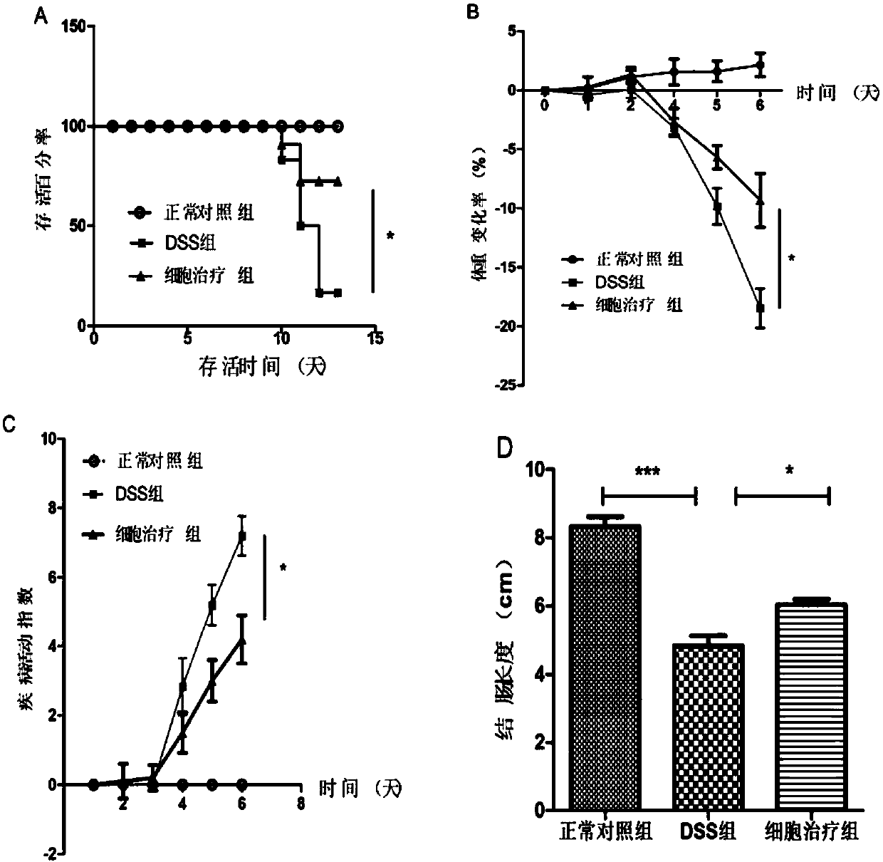
[0118] Embodiment 3 utilizes the preparation of embodiment 2 to treat the inflammatory bowel disease induced by mouse DSS
[0119] C57BL / 6 mice were adapted to the environment for 3 days, and were randomly divided into three groups, namely, the normal control group, the DSS model group (DSS is dextran sulfate sodium, which is a modeling reagent for enteritis model, purchased from MP Company) and the cell therapy group . The mice were given 3.5% DSS water for 6 days, and the cell therapy group injected 1x10 6 Individual Wharton area umbilical cord mesenchymal stem cell preparation (also known as MSCs), the injection volume is 100 microliters per mouse. In the DSS model group, PBS buffer solution with a volume of 100 microliters per rat was injected through the tail vein on the 1st, 3rd and 5th days of DSS water administration. The mice were weighed every day, and the degree of blood in stool and loose stool was detected. At the end of the experiment, the mice were sacrificed...
PUM
 Abstract
Description
Claims
Application Information