Combined diagnosis paper-base micro fluidic chip, and preparation method thereof
A microfluidic chip and paper-based technology, applied in the field of biomedical detection, can solve the problems of inability to achieve quantitative detection, long detection time, and complicated operation, and achieve the effects of saving detection time, low cost, and simplifying detection steps.
- Summary
- Abstract
- Description
- Claims
- Application Information
AI Technical Summary
Problems solved by technology
Method used
Image
Examples
Embodiment 1
[0044] 1. Quantum dot immunochromatography was used to detect C-reactive protein. The quantum dots were labeled on the CRP mouse monoclonal antibody and sprayed on the quantum dot reaction channel (3-3). At the same time, the CRP mouse monoclonal antibody was used as the detection line, and the goat anti-mouse IgG secondary antibody was used as the quality control line to coat the nitrocellulose membrane, and assembled with the absorbent pad and the optimized sample pad to form a test strip.
[0045] 2. Synthesis of quantum dot-labeled CRP antibody
[0046] Activation: Add the water-soluble quantum dots coated with carboxyl groups on the surface to the BS solution, add the mixed solution of NHS and BS and the solution of EDC and MES, vortex and mix, and activate by ultrasonic. Increase the volume, use a low-temperature ultracentrifuge for centrifugation, and discard the supernatant.
[0047] Coupling: Add BS solution to the activated quantum dots, reconstitute under vortex a...
Embodiment 2
[0049] Synthetic quantum dot-labeled PCT antibody
[0050] Water-soluble method of oil-soluble CdSe / ZnS quantum dots, or metal nanocluster quantum dots, or CdSe / ZnS quantum dots combined with metal nanoclusters: Precipitate with acetone and redisperse in chloroform, add an appropriate amount of mercapto Acetic acid, mix well and leave to react for 2h. Centrifuge, discard the supernatant, add buffer solution to dissolve completely, then add acetone to purify, repeat this process 2-3 times, and finally disperse the precipitate in phosphate buffer solution and store for later use.
[0051] The use of anti-anti-calcitonin monoclonal antibody and anti-calcitonin polyclonal antibody bound to two different sites of PCT, respectively, can basically rule out cross-reactivity. The monoclonal antibody is linked to the quantum dot surface by covalent cross-linking method to obtain the quantum dot-labeled monoclonal antibody. The specific process is: add quantum dots, EDC, 15 μg NHS solut...
Embodiment 3
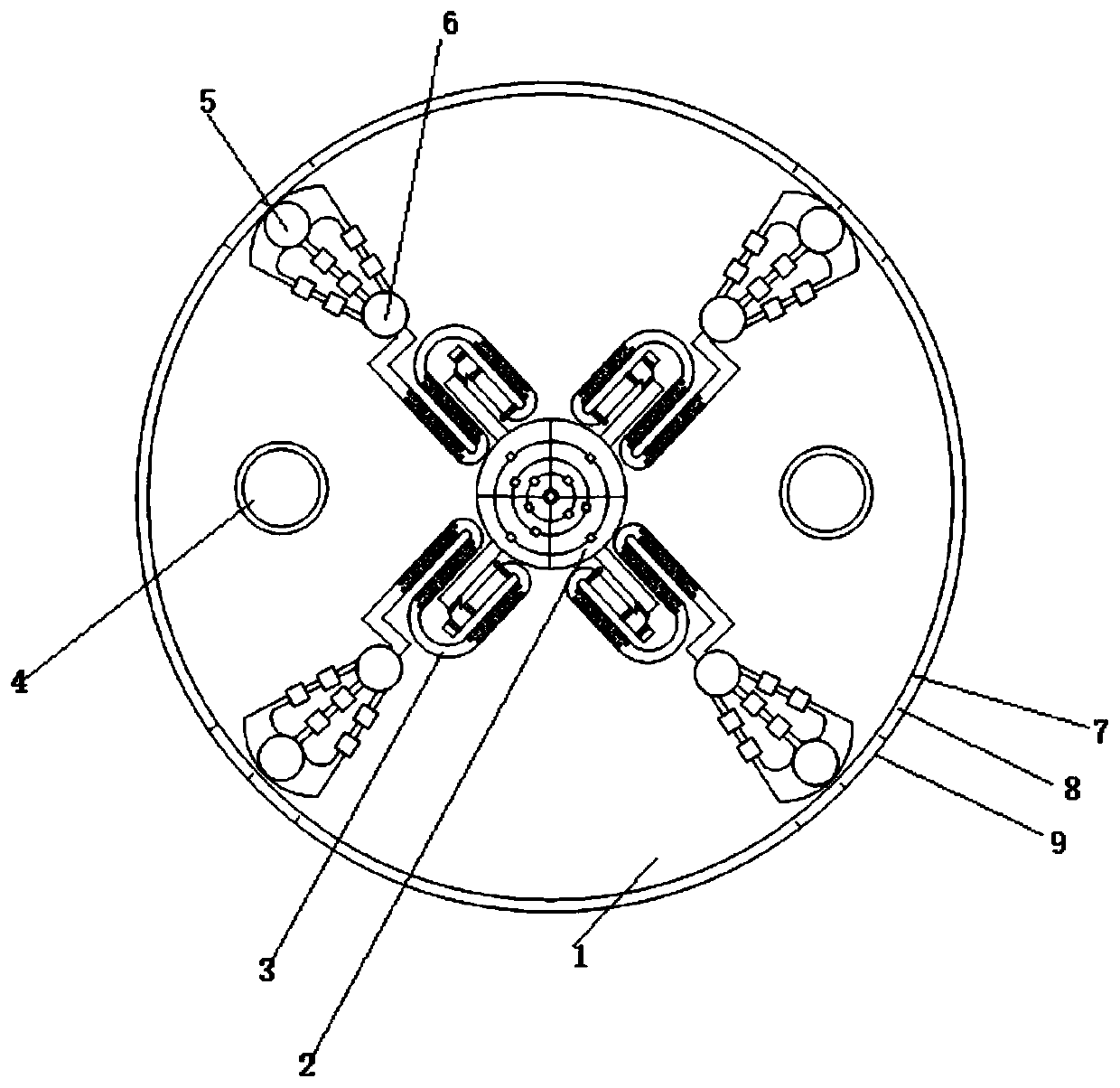
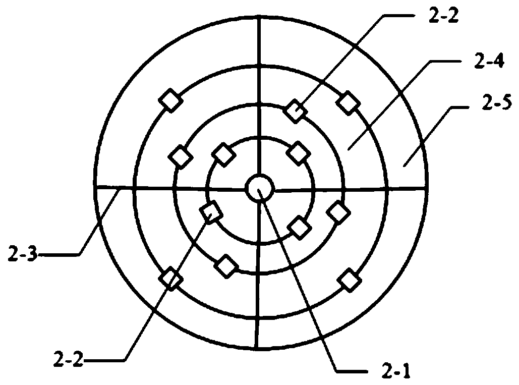
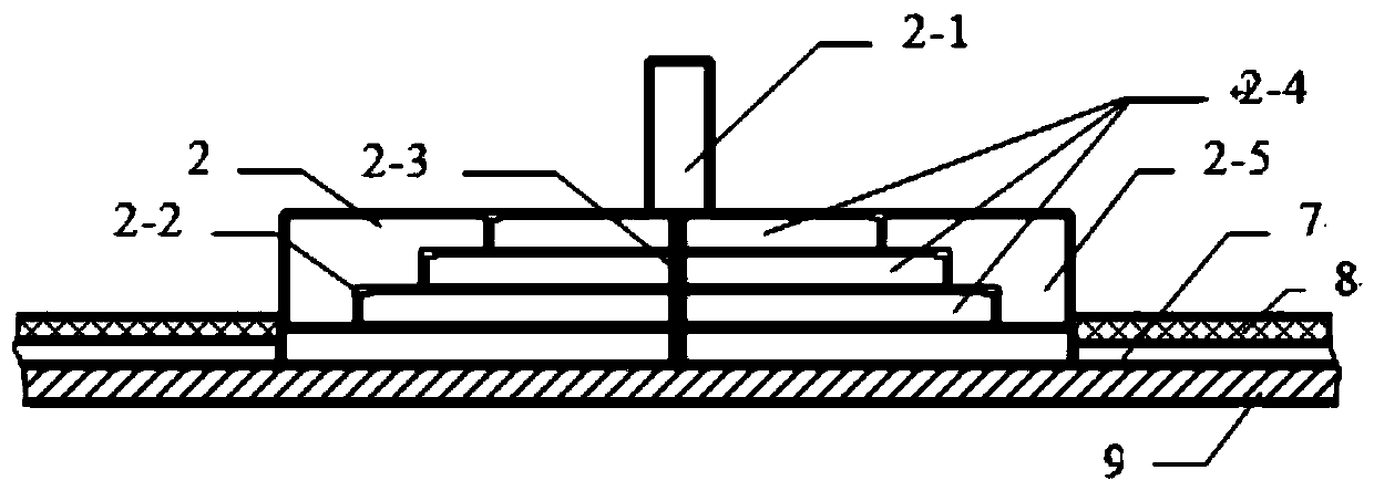
[0053] A combined diagnostic paper-based microfluidic chip: In one embodiment, the chip is mainly composed of various functional areas of the paper base (7), and the sample feeding part (2) and the sample dividing part (6). That is, the chip (1) is disc-shaped, with a paper base (7) and a chip substrate (9) on which the paper base (7) is placed, and a sealing film (8) on the paper base (7). Three layers; set a round hole at the center of the chip (1), place the sampling part (2) in the round hole, arrange more than 2 samples evenly and symmetrically along the diameter extension line around the sampling part (2) processing area (3); each sample processing area (3) is connected to a sample dividing part (6) at the outlet along the diameter extension line; each sample dividing part is connected to a detection area (5); More than 2 screw holes (4) are set along the direction of the extension of the diameter for fixing.
[0054] In one of the embodiments, the sampling part (2) is ...
PUM
 Abstract
Description
Claims
Application Information
 - R&D
- Intellectual Property
- Life Sciences
- Materials
- Tech Scout
- Unparalleled Data Quality
- Higher Quality Content
- 60% Fewer Hallucinations
Browse by: Latest US Patents, China's latest patents, Technical Efficacy Thesaurus, Application Domain, Technology Topic, Popular Technical Reports.
© 2025 PatSnap. All rights reserved.Legal|Privacy policy|Modern Slavery Act Transparency Statement|Sitemap|About US| Contact US: help@patsnap.com


