Combined nephroscope

A combined nephroscopic technology, applied in the field of nephroscopy, can solve the problems of operation failure, stone extraction channel loss, blind expansion, etc., and achieve the effect of reducing operation pressure and work intensity, fast operation time and reducing injury

Pending Publication Date: 2020-02-28
李文成
View PDF0 Cites 1 Cited by
  • Summary
  • Abstract
  • Description
  • Claims
  • Application Information

AI Technical Summary

Problems solved by technology

Each type of nephroscope can only complete stones with corresponding indications, and there is no universal nephroscope suitable for all surgical indications
[0004] 2. At present, all kinds of nephroscopes have only one channel, which is shared by the water inlet channel and the operation channel.
When ultra-fine nephroscopes (F7-8) and fine nephroscopes (F12-16) are used to operate on kidney and ureteral calculi with corresponding indications, due to the limitation of the diameter of the nephroscope, the water inlet and operation channel are relatively thin, and when broken When stone tools (holmium laser, ballistic probe) or lithograph forceps and other operating equipment enter the water inlet and operation channel, the water flow will be affected, resulting in unclear surgical field of view, affecting surgical safety and surgical efficiency
[0005] 3. At present, the function of the nephroscope is single, and it is only used for crushin

Method used


Image

  • Combined nephroscope
  • Combined nephroscope
  • Combined nephroscope

Examples


Embodiment approach 1

[0051] Such as Figure 3-Figure 7 , Figure 10 and Figure 11 As shown, the technical solution provided by this embodiment is as follows:

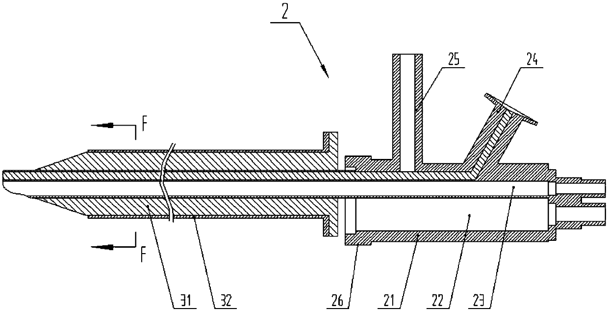
[0052] Includes core nephroscope 2 and dilator 3 for visual dilation;

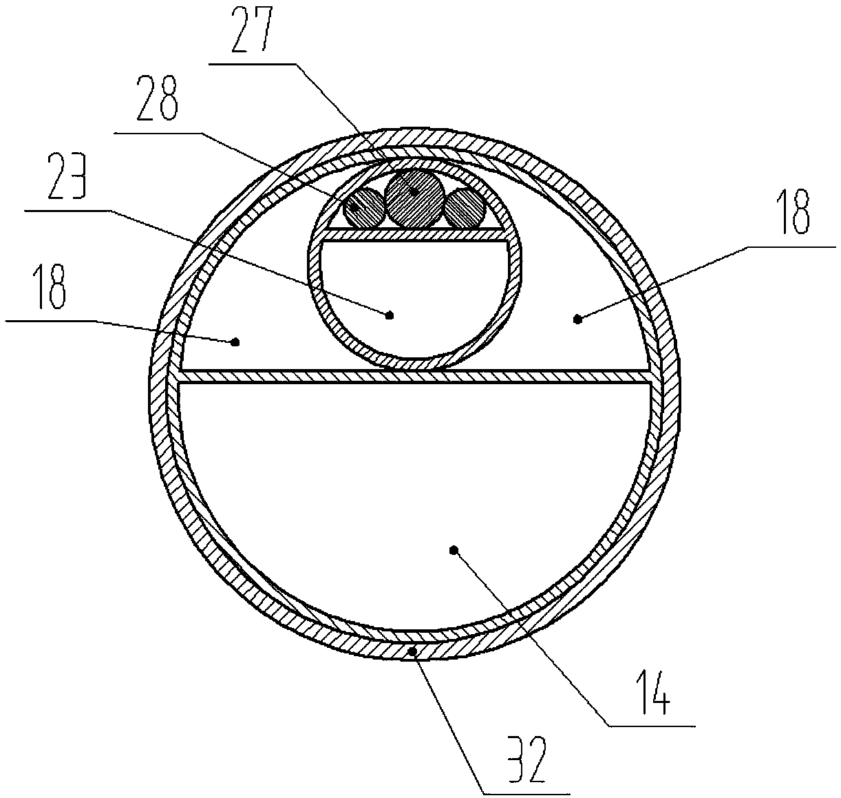
[0053] The core nephroscope 2 includes a camera system 27 and a light source system 28, and a first channel 23 for water inflow, water outflow and operation;

[0054] The dilator 3 is used for visual dilation after sheathed with the core nephroscope 2 , and includes a dilator inner core 31 and a dilator outer sheath 32 .

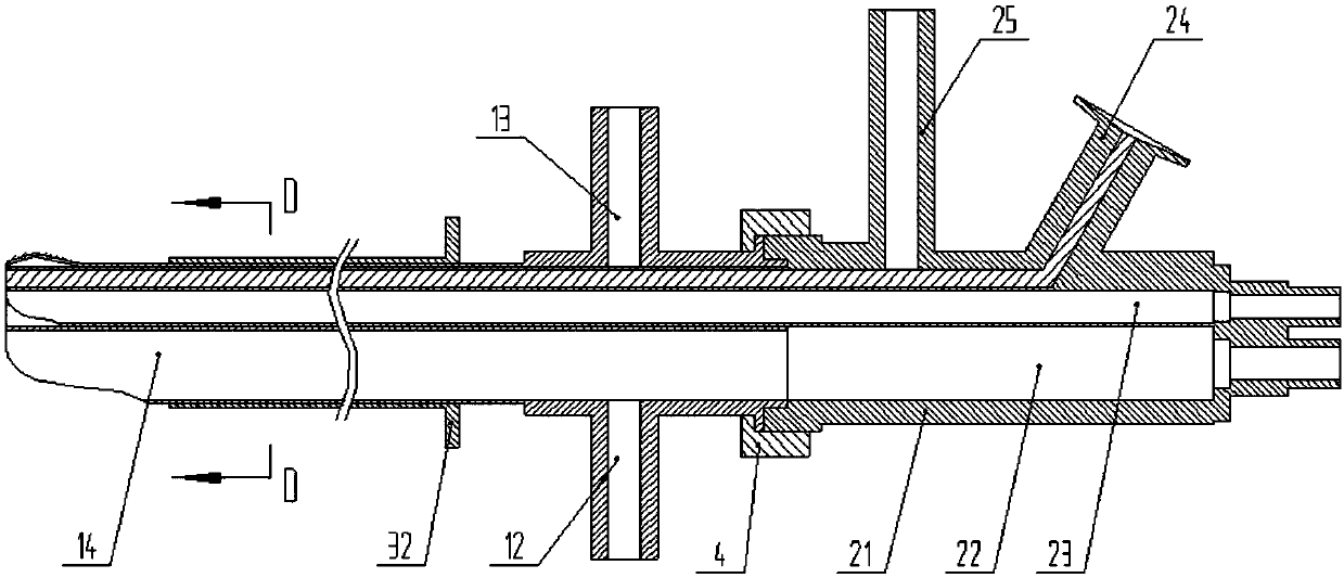
[0055] in, Figure 5-Figure 7 One of the structural methods of core nephroscope 2 is shown:

[0056] The core nephroscope 2 includes a hand-held end 21 and a working end. The working end is a slender rod, which is inserted into the body for surgical operations. The hand-held end 21 is outside the human body and is used for connecting various pipelines, observing and operating. The camera system 27 , the light source system 28 and the first channe...

Embodiment approach 2

[0072] Such as Figure 1-Figure 11 As shown, this embodiment, on the basis of Embodiment 1, adds a combined mirror sheath 1 that is detachably fitted and connected to the core nephroscope 2, and each preferred mode of Embodiment 1 can be combined with this embodiment .

[0073] Wherein, the combined mirror sheath 1 includes a nephroscope channel 15 for accommodating the core nephroscope 2, and a second operation channel 14 for water inlet, water outlet, negative pressure suction or mechanical operation;

[0074] The core nephroscope 2 is provided with a third operating channel 22 connected to the second operating channel 14. After the connecting, the second operating channel 14 and the third operating channel 22 form a straight line, which is convenient for instrument operation.

[0075] such as 8 and Figure 9 As shown, one of the structural modes of the combined mirror sheath 1 is shown:

[0076] The combined mirror sheath 1 also includes a working end and a hand-held end...


PUM


Abstract

The invention discloses a combined nephroscope which comprises a core nephroscope and an expander for visual expansion. The core nephroscope comprises a photographing system, a light source system anda first passage, and the expander is used for being sleeve-fitted with the core nephroscope for visual expansion and comprises an expander inner core and an expander outer sheath. By adopting the combined nephroscope, blind expansion in conventional art is completely avoided, and inserting depth of the expander and the core nephroscope can be observed visually, so that the combined nephroscope isenabled to accurately reach a calculous position in a kidney without going too shallow or too deep. Massive hemorrhage caused by damage to blood vessels of a patient is avoided completely, perforation is avoided, suffering of the patient is relieved, bodily damage to the patient is reduced, and the combined nephroscope is short in operative time, leads to little hemorrhage, small wound and quickrecovery of the patient and can be widely used in various kidney stone operations.

Description

technical field [0001] The invention relates to a surgical instrument, in particular to a nephroscope for checking, diagnosing and treating bladder or kidney. Background technique [0002] Percutaneous nephroscope lithotripsy (PCNL) is a modern minimally invasive technique for the treatment of kidney stones. Percutaneous nephroscope lithotripsy (PCNL) is to create a channel from the skin to the kidney in the waist, and insert the nephroscope through this channel. Kidney, using laser, ballistic, ultrasound and other lithotripsy tools to crush the kidney stones and take them out. But present nephroscope has following defect: [0003] 1. Currently, the existing nephroscopes are divided into ultra-fine nephroscopes (F7-8), thin nephroscopes (F12-16) and standard nephroscopes (F18-24) according to their diameters. Each type of nephroscope can only complete corresponding operations Indications, which type of nephroscope to use depends on the size and location of kidney stones an...

Claims


Application Information

Patent Timeline
IPC IPC(8): A61B1/307A61B1/32A61B17/34A61B17/02
CPCA61B1/00071A61B1/00105A61B1/307A61B1/32A61B17/0218A61B17/3403A61B2017/3413
Inventor 李文成章小平
Owner 李文成
Why Eureka
Try Eureka