A kind of physical specimen 3D printing method

A 3D printing and specimen technology, which is applied in the field of 3D printing of specimens, can solve problems such as the limitation of the production volume of plasticized specimens, achieve the effect of complete shape and structure, strong sense of reality, and solve inconvenience

Active Publication Date: 2022-03-15
河南中博科技有限公司
View PDF0 Cites 0 Cited by
  • Summary
  • Abstract
  • Description
  • Claims
  • Application Information

AI Technical Summary

Problems solved by technology

[0006] The purpose of the present invention is to provide a 3D printing method for physical specimens to solve the problem that the production volume of existing plasticized specimens is limited

Method used


Image

  • A kind of physical specimen 3D printing method
  • A kind of physical specimen 3D printing method
  • A kind of physical specimen 3D printing method

Examples


Embodiment 1

[0043] A kind of physical specimen 3D printing method, comprises the following steps:

[0044] (1) Antiseptic treatment: select a complete, non-trauma-free, and disease-free human specimen, and perform antiseptic treatment on the specimen.

[0045] Perfuse the specimen with formalin with a volume concentration of 15%, infiltrate the formalin into various parts of the specimen through the large blood vessels (femoral artery, common carotid artery), and finally put the formalin with a volume concentration of 10%. Soak in liquid for 3 months.

[0046] (2) Anatomical preparation, making the required specimens according to the anatomical atlas.

[0047] (2-1) Collect materials, cut the abdominal wall along the midaxillary line, cut off the inferior vena cava, hepatic portal vein, hepatic artery, common bile duct, and diaphragm with scissors, and take out the liver;

[0048] (2-2) Make, remove the superficial fascia on the liver and blood vessel wall, trim it neatly, keep 1 cm at ...

Embodiment 2

[0080] A kind of physical specimen 3D printing method, comprises the following steps:

[0081] (1) Antiseptic treatment: select human specimens that are complete, free from trauma, fractures, and lesions, and carry out antiseptic treatment on the human specimens.

[0082] Perfuse the specimen with formalin with a volume concentration of 20%, penetrate the formalin into various parts of the specimen through the large blood vessels (femoral artery, common carotid artery), and finally put the formalin with a volume concentration of 10%. Soak in liquid for 3 months.

[0083] (2) Anatomical preparation, making the required specimens according to the anatomical atlas.

[0084] (2-1) Collect materials, cut the abdominal wall along the midaxillary line, cut off the inferior vena cava, hepatic portal vein, hepatic artery, common bile duct, and diaphragm with scissors, and take out the liver;

[0085] (2-2) Make, remove the superficial fascia on the liver and blood vessel wall, trim i...

Embodiment 3

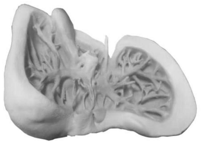
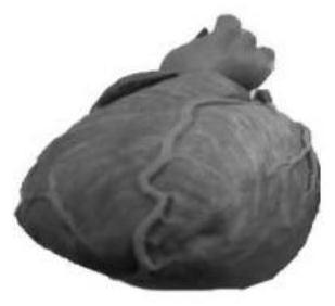
[0106] A method for 3D printing of physical specimens, the difference from Embodiment 1 is only in step (2). In this embodiment, human heart specimens are selected.

[0107] Anatomical fabrication includes:

[0108] (2-1) Material collection: Along the midline of the clavicles on both sides, use a rongeur to bite open the ribs, use a scalpel to incise the intercostal muscles to open the chest wall, open the pericardium, and use scissors to bluntly separate the pulmonary veins, pulmonary arteries, superior vena cava, and inferior vena cava. After the vena cava, brachiocephalic trunk, left subclavian artery, and left common carotid artery are separated, the blood vessels are cut off with a scalpel, and the heart is taken out;

[0109] (2-2) Production: Remove the superficial fascia on the heart and blood vessels, retain the myocardium and left coronary artery, right coronary artery, pulmonary vein, pulmonary artery, superior vena cava, inferior vena cava, brachiocephalic trunk, ...


PUM


Abstract

The invention relates to a 3D printing method for physical specimens, comprising the following steps: (1) antiseptic treatment, carrying out antiseptic treatment on selected physical specimens; (2) anatomical production, producing required specimens according to the anatomical atlas; (3) bleaching and rinsing , bleach the specimen, and then rinse it with tap water; (4) trimming, trimming the anatomically prepared specimen; (5) scanning, using 3D scanning tools to scan the above specimen; (6) specimen image processing, using a computer to The scanned image is repaired and synthesized; (7) Printing, using a 3D printer to print the specimen. The invention uses real biological specimens as raw materials to print specimens, the shape and structure are intact and unchanged, the sense of reality is strong, the process is simple, and the time is short; different materials can be used for mass production, which solves the inconvenience caused by the shortage of physical specimens to experimental teaching.

Description

technical field [0001] The present invention relates to the technical field of 3D printing of specimens, in particular to a method for 3D printing of physical specimens. Background technique [0002] At present, the commonly used cross-sectional specimens for teaching and learning in schools include: plasticized specimens, dipped specimens, and engraved mold-made model specimens. [0003] The engraving mold model specimen has a simple structure, but the size of the produced specimen is rigid, inflexible, complicated in process and high in cost. There is a certain deviation between the teaching and the real specimen, and the display of the name of the real structure of the specimen cannot be realized. [0004] The preparation process of soaked specimens is simple, they need to be preserved in the preservation solution, and they cannot be exposed to the air for a long time, and they are poisonous and have a pungent smell, so it is impossible to directly feel the position and ...

Claims


Application Information

Patent Timeline
Patent Type & Authority Patents(China)
IPC IPC(8): G09B25/00B29C64/386B33Y50/00G06T15/00G06T15/04G06T17/20G06T19/20
CPCG09B25/00B29C64/386B33Y50/00G06T15/005G06T15/04G06T17/20G06T19/20
Inventor 程明亮赵顺刘起颖赵文忠冯培勋贠跃进吉晓磊张帅斌
Owner 河南中博科技有限公司
Why Eureka
Try Eureka