PARP-1 single particle detection method based on dark field scattering imaging

A PARP-1, scattering imaging technology, applied in the biological field, can solve the problems of complex operation, long experimental process, and inability to detect PARP-1 activity, etc., and achieve the effect of high detection sensitivity

Active Publication Date: 2020-04-17
SOUTHEAST UNIV
View PDF9 Cites 4 Cited by
  • Summary
  • Abstract
  • Description
  • Claims
  • Application Information

AI Technical Summary

Problems solved by technology

Although these methods have high sensitivity, the experimental process is relatively long, the operation is relatively complicated, and radioactively labeled substrates are often required, so the detection cost is usually relatively high
[0004] A series of strategies for the detection of PARP-1 activity based on bulk measurements have been developed, including colorimetric, spectrofluorometric, electrochemical, and quartz crystal microbalance (QCM) methods, but these assays cannot detect PARP at the single-particle level -1 activity and imaging of PARP-1 in living cells, thus limited practical application

Method used


Image

  • PARP-1 single particle detection method based on dark field scattering imaging
  • PARP-1 single particle detection method based on dark field scattering imaging
  • PARP-1 single particle detection method based on dark field scattering imaging

Examples


Embodiment 1

[0059] Embodiment 1 diameter is the gold nanoparticle (Au 50 )Synthesis

[0060] First, gold nanoparticles (Au 13 ): Trisodium citrate solution (38.8mM, 5mL) was quickly added to stirring and boiling chloroauric acid solution (1mM, 50mL), the color of the mixed solution changed from yellow to colorless within 2min, and then turned into black and purple , finally turned into wine red, continue to stir and keep boiling for 15 minutes, after naturally cooling to room temperature, store in a 4°C refrigerator for later use;

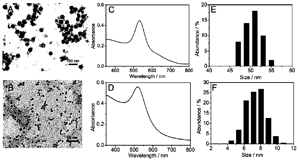
[0061] Secondly, gold nanoparticles (Au 50 ): Mix 25mL ultrapure water, 1mL freshly prepared Au 13 , NH 2 OH·HCl solution (0.2M, 360 μL) and polyvinylpyrrolidone solution (PVP, 1% w / v, 300 μL) were sequentially added to a 50 mL round bottom flask, and then 8 mL of 0.1 wt% chloroauric acid was added dropwise to the above mixture at room temperature solution and vigorously stirred for 30min, and left at room temperature for 30min after the stirring was stoppe...

Embodiment 2

[0063] Embodiment 2 diameter is the electropositive gold nanoparticle (Au 8 )Synthesis

[0064] First, chloroauric acid solution (1mM, 15mL) and CTAB (10mM, 2mL) were continuously stirred at room temperature for 15min; secondly, NaBH 4 (10mM, 2mL), the color of the solution changed from saffron yellow to orange red, and did not change within 15min, indicating that Au 8 , the obtained Au 8 Store in a brown glass bottle and store in a refrigerator at 4°C until use.

[0065] figure 2 Au is shown 8 sign of figure 2 B is Au 8 The TEM image is a uniform spherical shape with good monodispersity in solution; figure 2 D is Au 8 UV-Vis spectrum, the peak is concentrated at 518nm; figure 2 F is Au 8 The particle size distribution data, mainly distributed in 8nm. The above results indicate the successful synthesis of Au 8 .

Embodiment 3

[0066] Example 3 Principle Verification of Poly ADP-ribose Polymerase-1 Single Particle Detection Method Based on Dark Field Scattering Imaging

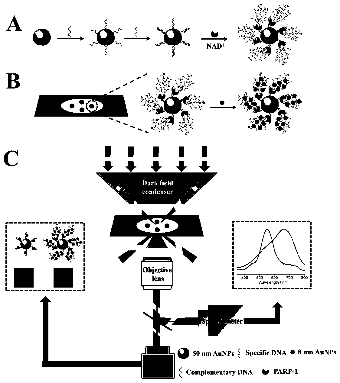
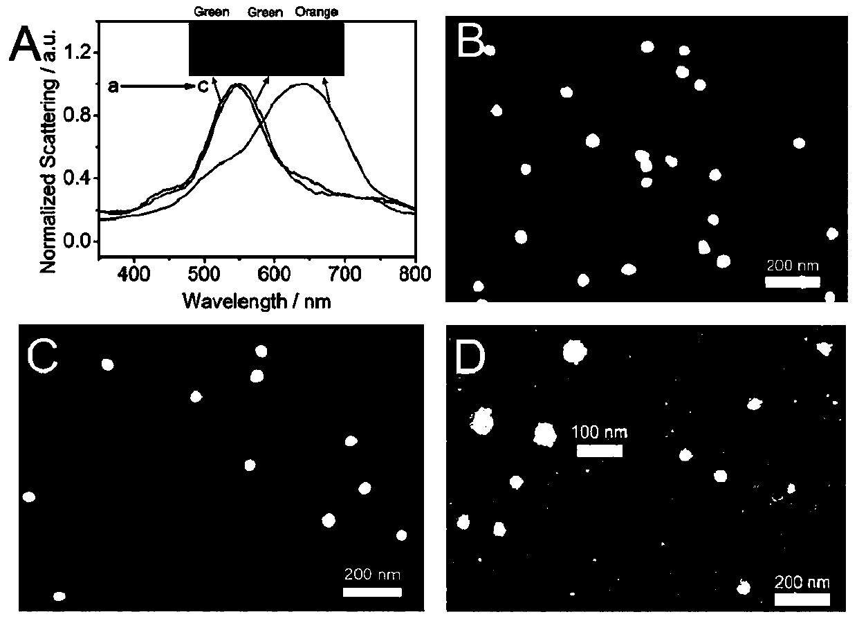
[0067] First, modify the two active DNA strands (s-DNA and c-DNA, see Table 1 for the sequence) to the Au prepared in Example 1 50 Au formed on the surface 50 -dsDNA, then add PARP-1, PARP-1 is Au 50 Active dsDNA on the surface activates the NAD + Cleavage into nicotinamide and ADP ribose, these units are linked to PARP-1 through covalent bonding and polymerized to form hyperbranched poly ADP ribose polymer (PAR), then in order to avoid agglomeration during the adsorption process, Au 50 -dsDNA@PAR is coated on the amino-functionalized glass for electrostatic adsorption for 5 minutes, the unadsorbed liquid is sucked away, and then Au is added 8 to form Au 50 -dsDNA@PAR@Au 8 . Compared with ds-DNA, PAR has more negative charges and can adsorb more Au 8 , so that Au 50 The scattering peaks were significantly red-shifted and exhi...


PUM

PropertyMeasurementUnit
particle diameteraaaaaaaaaa

Abstract

The invention discloses a polyadenosine diphosphate ribose polymerase-1 (PARP-1) single particle detection method based on dark field scattering imaging, and further discloses a label-free kit for identifying cancer cells. Quantitative detection of PARP-1 activity and dark field imaging of PARP-1 in cells on a single particle level are realized based on change of scattering spectral displacement of nanoparticles, so that cancer cells and normal cells are remarkably distinguished. White light is used as a light source to be combined with a dark field microscope and a single particle scatteringspectrometer, dark field imaging and scattered spectrum collection of a single plasma nanoparticle can be achieved, and compared with a traditional average measuring means, the method has higher detection sensitivity. The kit provided by the invention has the advantages of no labeling, high detection sensitivity, high spatial resolution and the like.

Description

technical field [0001] The invention relates to the field of biotechnology, in particular to a method for detecting single particles of poly ADP-ribose polymerase-1 (PARP-1) based on dark-field scattering imaging. Background technique [0002] Polyadenosine diphosphate-ribose polymerase-1 (PARP-1) is a multifunctional protein post-translational modification enzyme widely present in eukaryotic cells, involved in DNA replication and transcription, and maintaining genome stability. According to reports, PARP-1 is one of the members of the PARP family, which occupies a core function in the process of base excision repair and is a molecular sensor of DNA damage. When PARP-1 is activated, the activated PARP-1 can combine to form a homodimer and catalyze nicotinamide adenine dinucleotide (NAD + ) is decomposed into nicotine and ADP ribose, and poly ADP ribose polymer (PAR) is generated onto the target protein. Once the PAR with a large amount of negative charge on the surface is ...

Claims


Application Information

Patent Timeline
Patent Type & Authority Applications(China)
IPC IPC(8): G01N21/25
CPCG01N21/25
Inventor 卫伟张多多刘松琴
Owner SOUTHEAST UNIV
Why Eureka
Try Eureka