Kit for quantitatively detecting non-structural protein residues in foot-and-mouth disease inactivated antigen and application thereof

An inactivated antigen quantitative detection technology, applied in the biological field, can solve the problems of time-consuming and costly, and achieve the effect of high sensitivity and accurate calculation results

Inactive Publication Date: 2020-06-12
LANZHOU INST OF VETERINARY SCI CHINESE ACAD OF AGRI SCI
View PDF5 Cites 0 Cited by
  • Summary
  • Abstract
  • Description
  • Claims
  • Application Information

AI Technical Summary

Problems solved by technology

However, animal experiments are expensive and time-consuming. If a batch of vaccines cannot meet the purity requirements, the manufacturer must discard the entire batch of vaccines.
At pr

Method used


Image

  • Kit for quantitatively detecting non-structural protein residues in foot-and-mouth disease inactivated antigen and application thereof
  • Kit for quantitatively detecting non-structural protein residues in foot-and-mouth disease inactivated antigen and application thereof
  • Kit for quantitatively detecting non-structural protein residues in foot-and-mouth disease inactivated antigen and application thereof

Examples


Embodiment 1

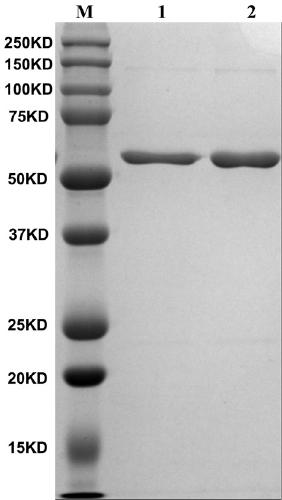
[0054] Example 1 Preparation of 3ABC monoclonal antibody and polyclonal antibody

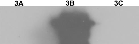
[0055] The foot-and-mouth disease NSP 3ABC of prokaryotic expression is purified ( figure 1 ), and perform gradient dialysis refolding on it, and then prepare monoclonal antibody by 3ABC immunized mice after refolding, subcloning the positive hybridoma cells three times by limiting dilution method, and screening out highly reactive monoclonal cells, Then the screened cells were injected into the peritoneal cavity of mice to prepare ascites. Specific operation process: Inject 0.5ml of Freund's incomplete adjuvant into the peritoneal cavity of female Balb / c mice, and inject about 7.0×10⁵ hybridoma cells into the peritoneal cavity 3 days later. After 9-10 days, ascitic fluid was collected and purified. Verification by Western blot showed that the monoclonal antibody recognized 3B protein, and WB results also showed that the monoclonal antibody recognized a linear epitope ( figure 2 ).

[0056]...

Embodiment 2

[0057] Example 2 Optimization of the CLIA method for detecting foot-and-mouth disease non-structural proteins and establishment of a standard curve

[0058] Dilute the 3ABC polyclonal antibody to 0.5 μg / ml as the capture antibody, dilute the 3B monoclonal antibody-HRP 1:10,000, 1:30,000, and 1:100,000 times as the detection antibody, and dilute the prokaryotic expressed foot-and-mouth disease NSP 3ABC to 500 with PBST, 250, 100, 50, 25, 10, 5, 2.5, 1, 0.5, 0.25, 0.1, 0.05, 0.025, 0.01, 0 ng / ml were used as standard antigens. Considering economic factors and detection range, the optimal coating concentration of capture antibody was finally determined to be 0.5 μg / ml, the optimal dilution of detection antibody was 1:10,000, and the optimal reaction time was 30 min, and a standard curve was established. Such as image 3 As shown, when the concentration of 3ABC is 0.1-10ng / ml, the measured chemiluminescence intensity has a linear relationship with the concentration of 3ABC, and t...

Embodiment 3

[0059] Embodiment 3 Determination of the optimal dilution factor of the foot-and-mouth disease inactivated antigen sample to be inspected

[0060] Since some antigens contain some components that affect the antigen-antibody reaction, the sample to be tested is diluted 1:2, 1:4, 1:8 and compared with the chemiluminescence value (CLIA value) of the original solution to determine the optimal dilution Spend. By comparing 8 antigens purified for different times (not one strain was used), it can be found that after serial dilution of some antigens, the CLIA value decreases proportionally (such as a, b, c, d); while some antigens are serially diluted After that, the CLIA value began to decrease proportionally from 1:4-1:8 (such as 1, 2); and the amount of NSPs contained in the antigen purified twice was already lower than the detection limit of this method, so after serial dilution, CLIA Values ​​vary little (eg I, II). Through this test, the optimal dilution of the antigen was fin...


PUM


Abstract

The invention relates to a chemiluminiscence kit for quantitatively detecting non-structural protein residues in foot-and-mouth disease inactivated antigens and application thereof, and belongs to thetechnical field of biology. The kit comprises a chemiluminiscence plate coated with a 3ABC polyclonal antibody, an enzyme-labeled monoclonal antibody, an antigen standard substance, a serum diluent,a 20 * PBST washing solution and a chemiluminiscence solution; the chemiluminiscence plate coated with the 3ABC polyclonal antibody is a white detachable polystyrene 96-pore plate; the enzyme-labeledmonoclonal antibody is an enzyme-labeled monoclonal antibody 9E2-HRP for recognizing 3B protein; the antigen standard substance contains 3ABC with the concentration of 10 ng/ml, BSA with the mass fraction of 0.2% and prolin 300 with the volume fraction of 0.05%. The kit disclosed by the invention is high in sensitivity, and can be used for quickly quantifying non-structural protein in the foot-and-mouth disease inactivated antigen.

Description

technical field [0001] The invention belongs to the field of biotechnology, and in particular relates to a kit for quantitatively detecting non-structural protein residues in inactivated antigens of foot-and-mouth disease and its application. Background technique [0002] Foot-and-mouth disease (FMD) is caused by foot-and-mouth disease virus (FMDV), an acute, febrile, highly contagious animal disease that infects cloven-hoofed animals such as pigs, cattle, and sheep. FMDV is a single-stranded positive-sense RNA virus belonging to the genus Foot-and-Mouth Disease Virus of the family Picornaviridae. 2A, 2B, 2C, 3A, 3B, 3C, 3D), and there are some uncleaved precursors such as 3ABC. The prevention and control of foot-and-mouth disease in my country mainly adopts comprehensive prevention and control measures of vaccine immunization, culling of infected and suspicious animals, and serum monitoring to control the epidemic of foot-and-mouth disease. In theory, only animals infecte...

Claims


Application Information

Patent Timeline
IPC IPC(8): G01N33/68G01N33/577G01N21/76
CPCG01N21/76G01N33/577G01N33/68
Inventor 常惠芸刘伟邵军军常艳燕
Owner LANZHOU INST OF VETERINARY SCI CHINESE ACAD OF AGRI SCI
Why Eureka
Try Eureka