Single cell mass spectrometry method
A mass spectrometry and single-cell technology, applied to the analysis of materials, individual particle analysis, particle and sedimentation analysis, etc., can solve the problems of reducing the convenience of operation, not conducive to popularization and application, and increasing sampling costs, so as to achieve convenient implementation and low cost. Low, easy-to-operate effect
- Summary
- Abstract
- Description
- Claims
- Application Information
AI Technical Summary
Problems solved by technology
Method used
Image
Examples
Embodiment 1
[0054] Lipid substance analysis in single MCF7 cell of embodiment 1
[0055] Cell fixation: the cultured MCF7 cells (7-8 μm) were transferred to PBS, centrifuged at 1200 rpm, and the cells were resuspended in phosphate buffered saline (PBS) containing 2.5% glutaraldehyde. After 20 minutes, Cells were collected by centrifugation and resuspended in water to obtain a cell suspension.
[0056] Preparation of capillary glass tube: use a P1000 needle puller to draw a capillary glass tube with a tip diameter of 5 μm.
[0057] Mass Spectrometry:
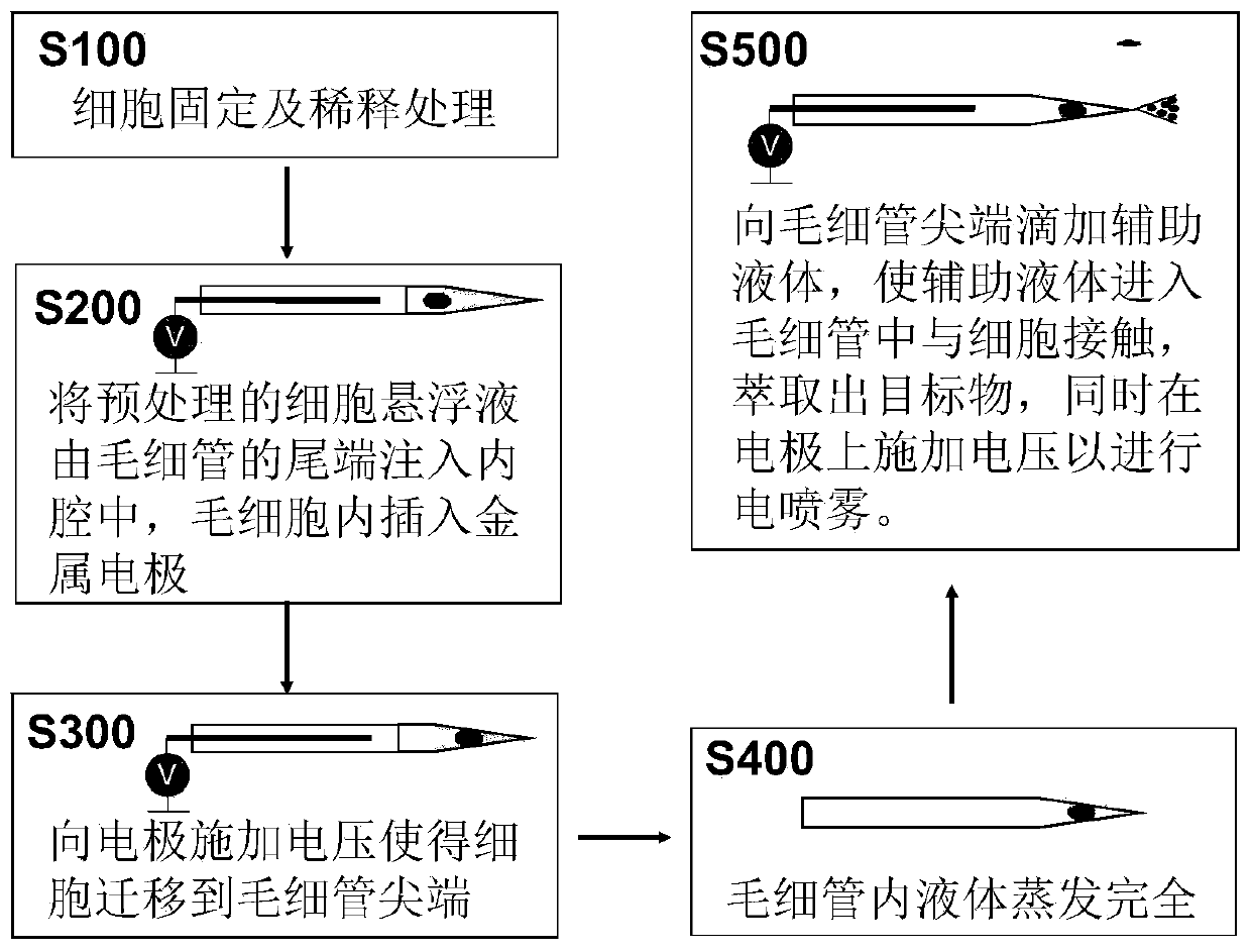
[0058] (1) Add 0.5 μL of cell suspension into the capillary glass tube from the end of the capillary glass tube, select the capillary glass tube containing 1 cell under the microscope, and use -1.2kV voltage to migrate a single MCF7 cell to the front of the capillary glass tube.
[0059] (2) Wait for the liquid in the capillary glass tube to evaporate completely.
[0060] (3) A voltage of -+1.8 kV is applied while dropping auxiliary liqui...
Embodiment 2
[0062] Lipid analysis of MCF7 cells after embodiment 2 single photochemical derivation
[0063] Cell fixation: transfer the cultured cells to PBS. Using 1200 rpm centrifugation, the cells were resuspended in phosphate buffered saline (PBS) containing 2.5% glutaraldehyde, and after 20 minutes, centrifuged, the cells were collected and resuspended in water.
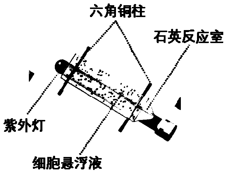
[0064] Photochemical derivation of cells: In the cell suspension, a solution of 2-acetylpyridine was added to a final concentration of 100 mM. In a quartz reaction box (such as figure 2 Shown) in, carry out ultraviolet photochemical reaction, used ultraviolet lamp is 254nm, and reaction time is 8 minutes.
[0065] Preparation of capillary glass tube: use a P1000 needle puller to draw a capillary glass tube with a tip diameter of 5 μm.
[0066] Mass Spectrometry:
[0067] (1) Add 0.5 μL of cell suspension (1-5 cells) into the capillary glass tube from the end of the capillary glass tube, select the capillary glass tube ...
Embodiment 3
[0072] Example 3 Structural identification of carbon-carbon double bond isomers of glycerophosphorylcholine (PC) in MCF7 cells after single photochemical derivatization
[0073] Cell fixation: transfer the cultured cells to PBS. Using 1200 rpm centrifugation, the cells were resuspended in phosphate buffered saline (PBS) containing 2.5% glutaraldehyde, and after 20 minutes, centrifuged, the cells were collected and resuspended in water.
[0074] Photochemical derivation of cells: In the cell suspension, a solution of 2-acetylpyridine was added to a final concentration of 100 mM. In a quartz reaction box (such as figure 2 Shown) in, carry out ultraviolet photochemical reaction, used ultraviolet lamp is 254nm, and reaction time is 8 minutes.
[0075] Preparation of capillary glass tube: use a P1000 needle puller to draw a capillary glass tube with a tip diameter of 5 μm.
[0076] Mass Spectrometry:
[0077] (1) Add 0.5 μL of cell suspension (1-5 cells) into the capillary gla...
PUM
| Property | Measurement | Unit |
|---|---|---|
| Outer diameter | aaaaa | aaaaa |
| Diameter | aaaaa | aaaaa |
Abstract
Description
Claims
Application Information
 - R&D
- Intellectual Property
- Life Sciences
- Materials
- Tech Scout
- Unparalleled Data Quality
- Higher Quality Content
- 60% Fewer Hallucinations
Browse by: Latest US Patents, China's latest patents, Technical Efficacy Thesaurus, Application Domain, Technology Topic, Popular Technical Reports.
© 2025 PatSnap. All rights reserved.Legal|Privacy policy|Modern Slavery Act Transparency Statement|Sitemap|About US| Contact US: help@patsnap.com


