
Preparation method of 19F magnetic resonance imaging nanoprobe

A technology of magnetic resonance imaging and nanoprobes, which is applied in the field of preparation of applied nanomaterials, can solve the problems of being unable to be directly applied to biological imaging, not being widely used, and having high biological toxicity, and achieves simple and fast preparation methods, low cost, Good biocompatibility effect

Inactive Publication Date: 2020-07-31
BEIJING UNIV OF CHEM TECH
View PDF1 Cites 2 Cited by
  • Summary
  • Abstract
  • Description
  • Claims
  • Application Information

AI Technical Summary

Problems solved by technology

[0003] However, currently 19 F MRI is not yet widely available
Because most fluorine-containing organic molecules are directly injected into the body with high biological toxicity, they cannot be directly applied to biological imaging
and commonly used in 19 The perfluorocrown ether molecules of F MRI are different. Although the cost of trifluoroethylamine is low, it contains highly active amino groups, which are easy to react with biomolecules in the body, high toxicity, and low boiling point. These characteristics make it impossible to use 19 F MRI

Method used


Image

  • Preparation method of 19F magnetic resonance imaging nanoprobe
  • Preparation method of 19F magnetic resonance imaging nanoprobe
  • Preparation method of 19F magnetic resonance imaging nanoprobe

Examples


Embodiment 1

[0018] a. Add 0.4g of polysuccinimide to 50mL of methanol, heat up and stir evenly, and put it into the reaction kettle;

[0019] b. Add 1.0mL trifluoroethylamine and 50mg amino PEG to the reaction kettle;

[0020] c. Put the reaction kettle into an oven and react at 200 degrees Celsius for 5 hours;

[0021] d. Take it out after cooling to room temperature, dry it in vacuum to remove the solvent, and the product obtained is dispersed in deionized water, and obtained by self-assembly 19 F Magnetic resonance imaging nanoprobes.

Embodiment 2

[0023] a. Add 0.4g polysuccinimide to 50mL ethanol, heat up and stir evenly, and put it into the reaction kettle;

[0024] b. Add 0.5mL trifluoroethylamine and 50mg amino PEG to the reaction kettle;

[0025] c. Put the reaction kettle into an oven and react at 200 degrees Celsius for 5 hours;

[0026] d. Take it out after cooling to room temperature, dry it in vacuum to remove the solvent, and the product obtained is dispersed in deionized water, and obtained by self-assembly 19 F Magnetic resonance imaging nanoprobes.

Embodiment 3

[0028] a. Add 0.4g of polysuccinimide to 50mL of N,N dimethylformamide, heat up and stir evenly, and put it into the reaction kettle;

[0029] b. Add 0.5mL trifluoroethylamine and 50mg amino PEG to the reaction kettle;

[0030] c. Put the reaction kettle into an oven and react at 200 degrees Celsius for 5 hours;

[0031] d. Take it out after cooling to room temperature, dry it in vacuum to remove the solvent, and the product obtained is dispersed in deionized water, and obtained by self-assembly 19 F Magnetic resonance imaging nanoprobes.


PUM


Abstract

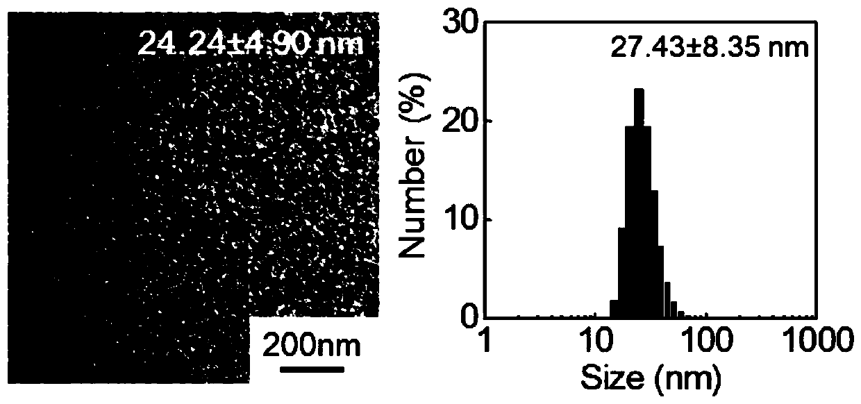
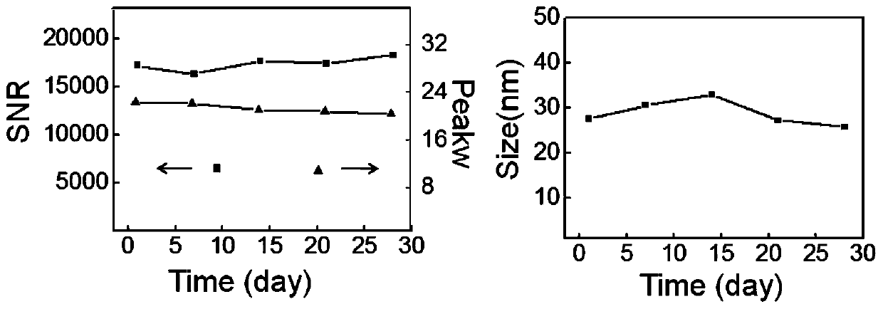
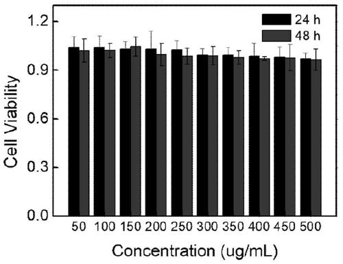
The invention discloses a preparation method of a 19F magnetic resonance imaging nanoprobe. The method comprises the following steps: in a polar organic solvent, carrying out an imide ring-opening reaction on polysuccinimide and trifluoroethylamine in a hydrothermal kettle with addition or no addition of amino-PEG, carrying out partial high-temperature carbonization, and then carrying out self-assembly to obtain the 19F magnetic resonance imaging nanoprobe. The probe has a good 19F NMR signal and can be directly used for 19F magnetic resonance imaging. The preparation method is simple and rapid and is low in cost, and the obtained nanoprobe is uniform in particle size distribution and good in biocompatibility, does not need hydrophilic modification, is high in stability and can be stored at normal temperature for a long time. The probe can attract wide attention in nano material science and biomedical science, and has important application prospects in biomedical fields such as biosensors, cell imaging, medical diagnosis and the like.

Description

technical field [0001] The invention belongs to the technical field of preparation of applied nanomaterials, in particular to a 19 F Magnetic resonance imaging nanoprobe preparation method. Background technique [0002] In recent years, magnetic resonance imaging (MRI) has been widely used in in vivo diagnosis because of its non-invasiveness and its ability to provide effective information on biological deep tissues without using radioactivity. so far, 1 H MRI has been widely used in clinical diagnosis. However, the commonly used 1 H MRI contrast agents usually contain gadolinium, which is likely to cause safety hazards. compared to 1 HMRI, 19 The F MRI signal has a chemical shift range of about 400ppm, which is very sensitive to changes in chemical bonds and microenvironment. The endogenous fluorine atoms in the human body only exist in the form of solid salts such as teeth and bones, which have no magnetic resonance signals and almost no background interference. ...

Claims


Application Information

Patent Timeline
Patent Type & Authority Applications(China)
IPC IPC(8): A61K49/12A61K49/18
CPCA61K49/12A61K49/1818
Inventor 汪乐余郭唱
Owner BEIJING UNIV OF CHEM TECH
Learn More