
Renal ureter fistulization urine shunt monitoring system

A monitoring system and ureter technology, applied in the field of medical devices, can solve the problems of working environment restrictions, damage to kidney function, excessive fluid accumulation, etc., and achieve the effect of improving flexibility, high stability, and not easy to fall off

Active Publication Date: 2020-08-07
SHANGHAI FIRST PEOPLES HOSPITAL
View PDF15 Cites 2 Cited by
  • Summary
  • Abstract
  • Description
  • Claims
  • Application Information

AI Technical Summary

Problems solved by technology

[0003] At present, after nephroureterostomy in clinical practice, if it is unilateral ureterostomy, the urine volume can be recorded unilaterally, but if it is bilateral unilateral ureterostomy, both ureteral stents will urinate in the ostomy bag at the same time, so it is impossible Record the urine output of both kidneys separately, and the ureteral stent is blocked by urine crystal precipitation and infection, which damages renal function. However, if the contralateral kidney urinates well, it often conceals the condition; It is very inconvenient to urinate even if the ostomy bag is full if there is a need for records; moreover, for urine testing, the latest urine is often needed to meet the testing requirements, and the existing ostomy drainage tubes do not Does not have the function of storing fresh urine
[0004] Chinese patent document: CN109646790A discloses a double J tube structure, including a tube body and a detection device fixed on the inner wall of the tube for detecting the flow rate and concentration of urine in the tube, and the detection device is wirelessly connected to an external terminal. After detecting the urine flow rate and concentration, the flow rate signal and concentration signal are output to the external terminal. The detection device includes a flow rate detector, a concentration detector and a wireless transmission board attached to the inner wall of the tube. The wireless transmission board Coupled with the analysis server, the flow rate detector and the concentration detector are wirelessly connected to the wireless transmission board. The advantage is that the flow rate and concentration of the urine in the tube can be effectively detected by setting the detection device in the tube. , which can effectively output the flow rate signal and concentration signal to the external terminal. After analysis and processing by the external terminal, the function monitoring of the patient's kidney and ureter can be realized, but it does not have the function of storing urine for testing, and its It is used to replace the ureter in the body, and its working environment has certain restrictions on it, and it does not have the replacement function
Its beneficial effect: the cleaning speed of the internal accumulation is fast, and the inside of the ostomy bag can be cleaned to ensure the cleanliness of the ostomy bag and avoid wound infection. , can store a lot of effusion; but it does not involve the function of separate statistics of the flow of each pipeline, so it does not solve the above problems

Method used


Image

  • Renal ureter fistulization urine shunt monitoring system
  • Renal ureter fistulization urine shunt monitoring system
  • Renal ureter fistulization urine shunt monitoring system

Examples


Embodiment 1

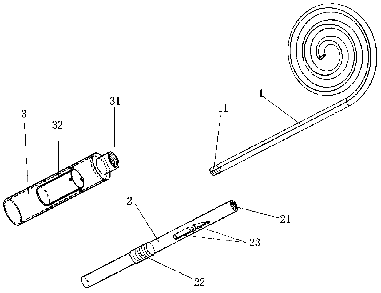
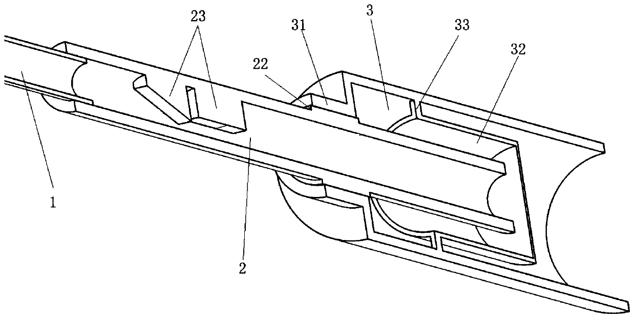
[0037] Please see attached figure 1 ; attached figure 1 It is a schematic diagram of the components of a nephroureterostomy urine diversion monitoring system of the present invention. A urine shunt monitoring system for nephroureterostomy, comprising a ostomy tube 1, a detection tube 2, and a liquid storage tube 3; the ostomy tube 1 is a tubular cavity structure with openings at both ends, and is a single J-tube structure, the The straight tube end of the fistula tube 1 is provided with a first connecting part 11, and the first connecting part 11 is an external thread structure; please refer to the attached figure 2 ; attached figure 2 It is a cooperating cross-sectional view of the detection tube and the liquid storage tube of a nephroureterostomy urine diversion monitoring system of the present invention. The detection tube 2 is a tubular cavity structure with openings at both ends, and one end is provided with a second connecting part 21, and the second connecting part...

Embodiment 2

[0047] Please see attached Figure 6 ; attached Figure 6 It is a sectional view of the liquid storage tube of another nephroureterostomy urine diversion monitoring system of the present invention. This embodiment is basically the same as Embodiment 1, the difference is that the liquid storage tube 3 includes a first sleeve 34 and a second sleeve 35, and the first sleeve 34 and the second sleeve 35 pass through Threads are detachably connected, the fourth connecting portion 31 is arranged on the first sleeve 34, the liquid storage tank 32 is fixed inside the second sleeve 35, and the open end of the liquid storage tank 32 is It is flush with the opening of the upper end of the second sleeve 35 . It should be noted that the two-stage arrangement of the liquid storage tube 3 does not affect the practical performance of the present invention, and a detachable connection structure is provided at the liquid storage tank 32, which can facilitate the taking of urine assay.


PUM


Abstract

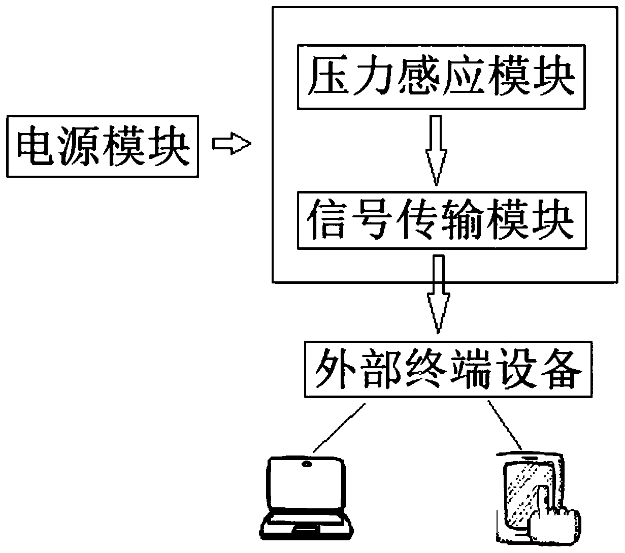
The invention relates to a renal ureter fistulization urine shunt monitoring system which comprises a fistulization tube, a detection tube and a liquid storage tube. One end of the fistulization tubeis used for being connected with a renal ureter, and the other end of the fistulization tube is provided with a first connecting part; the detection tube has a tubular cavity structure with openings in the two ends and is detachably connected with the fistulization tube. A flow monitoring device is arranged in the detection tube, comprises a pressure sensing module, a signal transmission module and a power supply module, and is used for monitoring the flow of liquid flowing through the detection tube; the liquid storage tube has a tubular cavity structure with openings in the two ends and is detachably connected with the detection tube, a liquid storage tank is arranged in the liquid storage tube, and the liquid storage tank can store liquid flowing out of the liquid storage tube. The renal ureter fistulization urine shunt monitoring system has the advantages that the urine volumes of the kidneys on the two sides are recorded respectively, the situation that the problem of the kidneyson the single side is covered is prevented, the function of storing latest urine for testing is achieved, and the stability and flexibility are high.

Description

technical field [0001] The invention relates to the technical field of medical devices, in particular to a urine diversion monitoring system for nephroureterostomy. Background technique [0002] Nephroureterostomy is a common urinary diversion in urology, which may be accompanied by self-scheduled total cystectomy, acute urinary tract obstruction, bilateral ureteral stones, or pyonephrosis, renal tuberculosis, etc. [0003] At present, after nephroureterostomy in clinical practice, if it is unilateral ureterostomy, the urine volume can be recorded unilaterally, but if it is bilateral unilateral ureterostomy, both ureteral stents will urinate in the ostomy bag at the same time, so it is impossible Record the urine output of both kidneys separately, and the ureteral stent is blocked by urine crystal precipitation and infection, which damages renal function. However, if the contralateral kidney urinates well, it often conceals the condition; It is very inconvenient to urinate ...

Claims


Application Information

Patent Timeline
IPC IPC(8): A61M1/00A61B5/20
CPCA61M1/0023A61B5/201A61B5/208A61M2205/3331A61M2205/3379A61M2210/1078A61M1/84A61M2210/005
Inventor 伍科刘志宏郑军华王翔李明清郑重姚智显周文洁郑心怡刘语瞳穆星宇孙丰王心怡凌静
Owner SHANGHAI FIRST PEOPLES HOSPITAL
Learn More