
A method for assessing the motor representation area of ​​the cerebral cortex of the trunk muscles and its application

A cerebral cortex, exercise technology, applied in applications, sports accessories, muscle training equipment, etc., to achieve the effect of clear and clear experimental results, easy to understand, and save the experimental site

Active Publication Date: 2022-01-14
THE FIRST AFFILIATED HOSPITAL OF SUN YAT SEN UNIV
View PDF3 Cites 0 Cited by
  • Summary
  • Abstract
  • Description
  • Claims
  • Application Information

AI Technical Summary

Problems solved by technology

The brain activates the transversus abdominis and multifidus muscles via corticospinal pathways that project to spinal motoneurons, however, this pathway is deep and cannot be achieved in vitro

Method used


Image

  • A method for assessing the motor representation area of ​​the cerebral cortex of the trunk muscles and its application
  • A method for assessing the motor representation area of ​​the cerebral cortex of the trunk muscles and its application
  • A method for assessing the motor representation area of ​​the cerebral cortex of the trunk muscles and its application

Examples


Embodiment 1

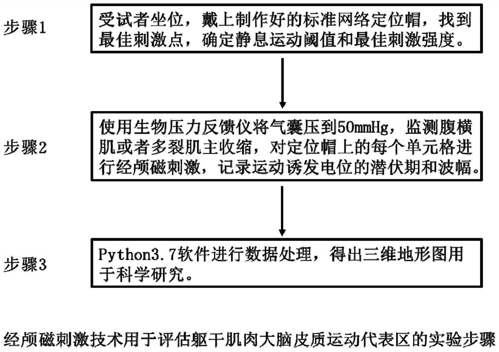
[0031] A kind of embodiment of the method for assessing trunk muscle cerebral cortex motor representative area of ​​the present invention, comprises the following steps (flow process is as follows figure 1 shown):

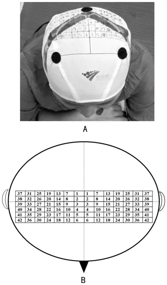
[0032] (1) The subject sits and wears a prepared standard network positioning cap (such as figure 2 ), find the best stimulation point, determine the resting motion threshold and the best stimulation intensity;

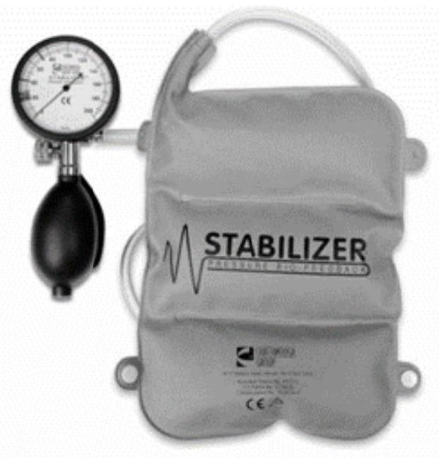
[0033] (2) Instruct the subject to use the biostress feedback instrument (such as image 3 ) to 50±2mmHg to make the transversus abdominis or multifidus complete about 10% of the maximum voluntary isometric contraction, perform transcranial magnetic stimulation on each 1×1cm cell on the positioning cap, and stimulate each grid 10 times, 5 out of 10 times the motor-evoked potential amplitude ≥ 50μV was considered as effective stimulation, and the latency and amplitude of the motor-evoked potential of effective stimulation were recorded;

[0034] (3) Pro...

Embodiment 2

[0039] The test-retest reliability of embodiment 2 biological pressure feedback instrument

[0040] Subjects: This study recruited 31 healthy young volunteers from our hospital interns, including 6 males and 25 females, aged 22.23±1.96 years old, weighing 56.65±8.98kg, height 164.19±7.54cm, BMI 21.00±2.59 kg / cm 2 . Inclusion criteria: ①BMI is ±20% standard; ②Right-handed; ③No musculoskeletal system disease, nervous system disease or mental and psychological disease; ④No history of abdominal, lumbar / sacral trauma, or surgery; ⑤No scoliosis ; ⑥ No muscle fatigue and low back pain in the past 6 months, and no strenuous exercise within 24 hours before the experiment. Exclusion criteria: ① Those who cannot complete the action tasks correctly; ② Female menstrual period; ③ Pregnant women. This experiment was approved by the Ethics Committee of our hospital, and all subjects signed informed consent.

[0041] Laboratory equipment:

[0042]The Pressure Biofeedback Unit (PBU) (Chatt...


PUM


Abstract

The invention discloses a method for evaluating the cerebral cortex motor representative area of ​​the trunk muscle and its application. The method monitors the contraction degree of the trunk muscle of the subject through a biological pressure feedback instrument, and simultaneously uses the transcranial magnetic stimulation technology to stimulate the cerebral cortex motor representative area. In the area, the amplitude and latency of the motor evoked potential of the trunk muscles were recorded, and finally the data was processed by Python3.7 software to obtain a three-dimensional image of the trunk muscles in the motor representative area of ​​the cerebral cortex. The method of the invention improves the large and bulky surface electromyography equipment into an easy-to-carry, convenient and compact biological pressure feedback instrument, simplifies the experimental equipment, saves the experimental site, makes the experimental process smoother, and has wider applicability. At the same time, using the programming software to effectively process the experimental results and generate three-dimensional images makes the experimental results more clear and vivid, which is of great significance for a better understanding of the relationship between pain-brain-muscle.

Description

technical field [0001] The invention relates to a method for evaluating the motor representative area of ​​the cerebral cortex of the trunk muscle and its application. Background technique [0002] Transcranial magnetic stimulation technology is a neurophysiological stimulation technology proposed by Barker et al. in 1985. Its basic principle is to generate a magnetic field in a coil through an electric current. The magnetic field can pass through the complete skull and generate local microcurrents in the corresponding cerebral cortex. These The current is sufficient to cause depolarization of the cerebral cortex and subcortical neurons, which can be used to study the integrity of the motor cortex physiology and motor system, including the basic mechanism of motor control and neuroplasticity under normal and pathological conditions. It is a non-invasive Means; Compared with other equipment such as functional magnetic resonance imaging, it is not limited by body position and ...

Claims


Application Information

Patent Timeline
Patent Type & Authority Patents(China)
IPC IPC(8): A61B5/389A61B5/377A61B5/383A61B5/22G06T15/00
CPCA61B5/7235A61B5/224A61B5/227G06T15/00A61B5/377A61B5/389
Inventor 李鑫王楚怀
Owner THE FIRST AFFILIATED HOSPITAL OF SUN YAT SEN UNIV
Learn More