Microscopic imaging method for simultaneously obtaining light absorption contrast and light scattering contrast

A microscopic imaging and double-contrast technology, which is applied in the fields of scattering characteristic measurement, color/spectral characteristic measurement, material analysis through optical means, etc., can solve the problem that it is difficult for optical microscopes to achieve clear imaging

Active Publication Date: 2020-09-22
NANJING UNIV
View PDF7 Cites 1 Cited by
  • Summary
  • Abstract
  • Description
  • Claims
  • Application Information

AI Technical Summary

Problems solved by technology

[0002] For transparent substances, such as most transparent cells, histopathological sections, etc., when light passes through these substances, the light intensity changes slightly, and it is difficult to achieve clear imaging with ordinary opti

Method used


Image

  • Microscopic imaging method for simultaneously obtaining light absorption contrast and light scattering contrast
  • Microscopic imaging method for simultaneously obtaining light absorption contrast and light scattering contrast
  • Microscopic imaging method for simultaneously obtaining light absorption contrast and light scattering contrast

Examples


Embodiment 1

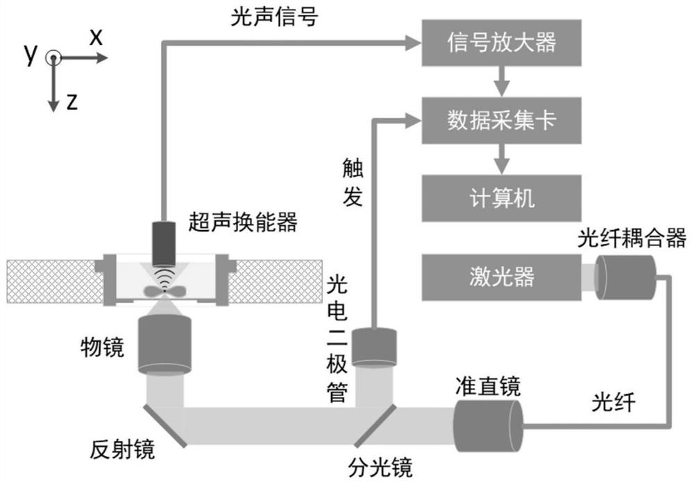
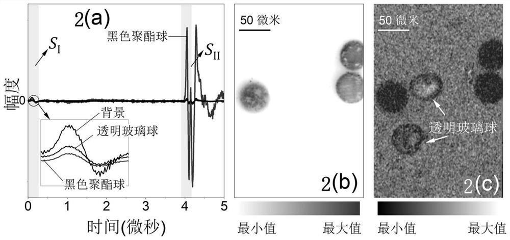
[0034] use figure 1 The system images binary mixed particles with different optical properties, including the following steps:

[0035] Step 1: The binary mixed particles composed of 50 micron diameter black polyester spheres and transparent glass spheres are spread on the bottom of the confocal dish, and distilled water is injected into the confocal dish to achieve ultrasonic coupling.

[0036] Step 2: The pulsed laser is coupled into the single-mode fiber through the fiber coupler. The output light of the single-mode fiber is collimated by the collimator and then enters the inverted microscope objective lens, and is focused on the sample through the bottom of the confocal dish. The numerical aperture of the inverted microscope objective lens is 0.6, the laser wavelength is 532nm, the pulse width is about 8ns, and the pulse repetition frequency is 10kHz. The photodiode is used to monitor the intensity of the laser and trigger the data acquisition card to collect the signal. ...

Embodiment 2

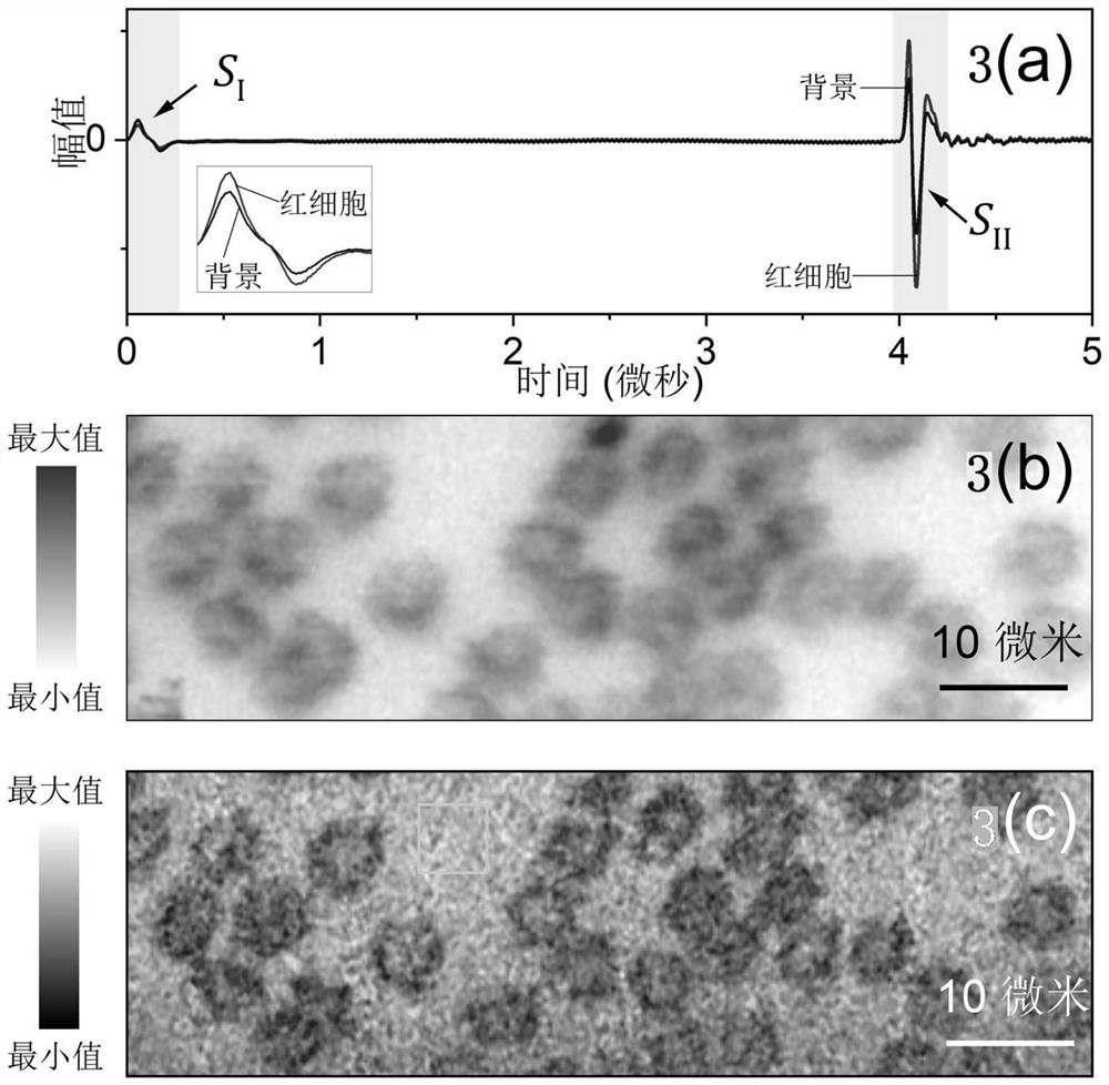
[0042] use figure 1 The system performs label-free imaging of red blood cells, including the following steps:

[0043] Step 1: Take fresh blood from a mouse (C57BL / 6JGpt, male, 10 weeks old, weighing about 25g), smear and fix it on the upper surface of the confocal dish, make a monolayer red blood cell smear, and inject it into the confocal dish Physiological saline for ultrasound coupling.

[0044] Step 2: The pulsed laser is coupled into the single-mode fiber through the fiber coupler. The output light of the single-mode fiber is collimated by the collimator and then enters the inverted microscope objective lens, and is focused on the sample through the bottom of the confocal dish. The numerical aperture of the inverted microscope objective lens is 0.6, the laser wavelength is 532nm, the pulse width is about 8ns, and the pulse repetition frequency is 10kHz. The photodiode is used to monitor the intensity of the laser and trigger the data acquisition card to collect the sig...


PUM


Abstract

The invention discloses a microscopic imaging method for simultaneously obtaining light absorption contrast and light scattering contrast. The method comprises the following steps: focusing pulse laser on a sample, absorbing part of the pulse laser by the sample, exciting an ultrasonic signal due to a photoacoustic effect, and receiving the ultrasonic signal through an ultrasonic transducer whichis confocal with a microscope objective and coaxially arranged to obtain an image with light absorption contrast; and meanwhile, using residual pulse laser penetrating through the sample irradiates the surface of the same ultrasonic transducer after being scattered and absorbed by the sample, so as to excite another ultrasonic signal, receiving the ultrasonic signal by the ultrasonic transducer, and obtaining an image with light scattering contrast. According to the invention, for any imaging point on the sample, light scattering-light absorption bimodal microimaging can be realized at the same time only by one-time light excitation; moreover, the method is a high-resolution imaging technology without an exogenous marker, avoids possible influence of a marker on the properties of the sample, and has higher usability and practicability.

Description

technical field [0001] The invention relates to a microscopic imaging method capable of simultaneously obtaining double-contrast ratios of light absorption and light scattering. Through one scan, microscopic images of two contrast ratios of sample light absorption and light scattering can be obtained simultaneously. Background technique [0002] For transparent substances, such as most transparent cells, histopathological sections, etc., when the light intensity changes slightly after passing through these substances, it is difficult to achieve clear imaging with ordinary optical microscopes. Therefore, staining is often required for imaging transparent substances such as living cells and histopathological sections, and label-free imaging is still an important challenge for the imaging of cells and histopathological sections. [0003] Light scattering is an effective imaging parameter for characterizing transparent substances such as cells. By measuring the intensity, phase...

Claims


Application Information

Patent Timeline
IPC IPC(8): G01N21/17G01N21/31G01N21/51
CPCG01N21/1702G01N21/31G01N21/51G01N2021/1734
Inventor 陶超吴诗晴刘晓峻
Owner NANJING UNIV
Why Eureka
Try Eureka