Carcino-embryonic antigen detection method
A carcinoembryonic antigen and detection method technology, applied in the field of analytical chemistry, can solve the problems of high background fluorescence and low sensitivity
- Summary
- Abstract
- Description
- Claims
- Application Information
AI Technical Summary
Problems solved by technology
Method used
Image
Examples
Embodiment 1
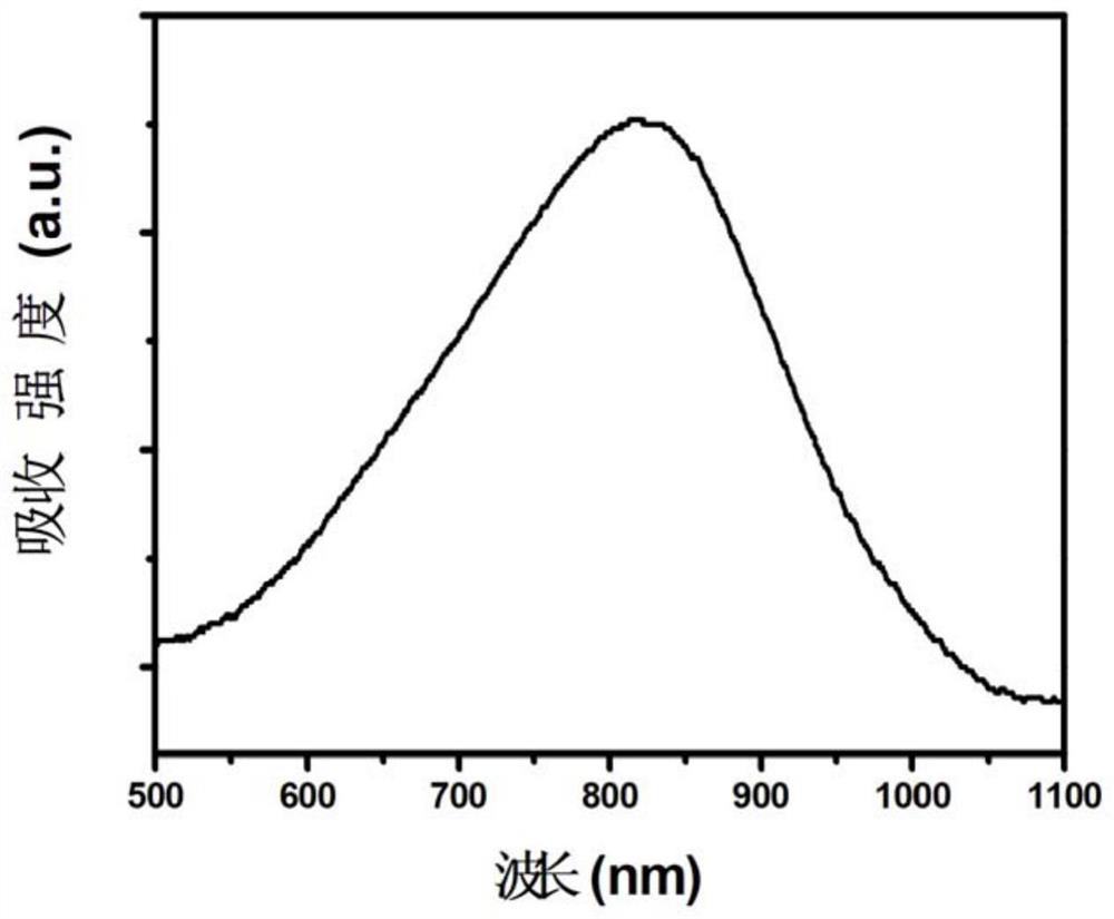
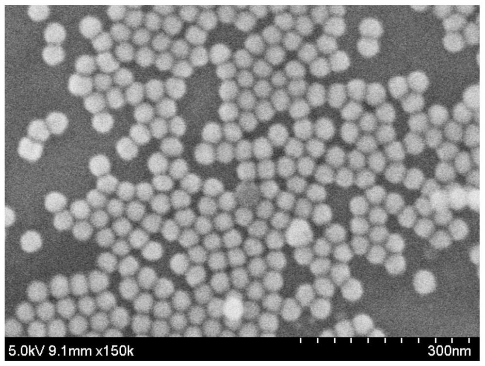
[0033] 1) Preparation of silver triangle nanoparticles: get 100mL deionized water, add 1mL 10mM silver nitrate, 1mL 15mM sodium citrate, 4mL 1.75mM PVP (polyvinylpyrrolidone) and 240 μL 30wt% hydrogen peroxide, and at room temperature Stir vigorously, then quickly add 1.2 mL of 100 mM NaBH 4 , the solution turned blue after about 30 minutes, and silver triangular nanoparticles were obtained. Detect the 500-1100nm absorption spectrum of the silver triangular nanoparticle solution, see the results figure 1 . From figure 1 It can be seen that silver triangular nanoparticles have been successfully prepared, and their absorption peaks are between 800 and 900 nm, which can be used to quench Nd 3+ Ions emit light at 840-900nm.
[0034] 2) Label the nucleic acid aptamer onto the silver triangle nanoparticles: Take 10 mL of the silver triangle nanoparticle solution obtained in step 1) and centrifuge twice at 10,000 rpm for 15 minutes each time, discard the supernatant and finally d...
Embodiment 2
[0037] The detection of embodiment 2 carcinoembryonic antigen
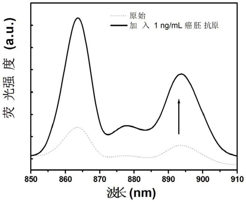
[0038] The silver triangular nanoparticle solution (1mL, 0.4nM) of the nucleic acid aptamer label obtained in Example 1 and the nucleic acid aptamer label Nd 3+ Ion-doped rare earth nanoparticles (1mL, 4nM) and carcinoembryonic antigen nucleic acid aptamer (50μL, 100nM, its nucleotide sequence is shown in SEQ ID NO.3) were mixed; after 30 minutes, CEA was added to make The final concentration of CEA in the mixed solution was 1 ng / mL CEA. Gently oscillate to mix, and incubate at constant temperature for 1 hour before testing. No CEA is added to the system as a control. Excited by a near-infrared wavelength of 808nm laser, scanning the near-infrared emitted light in the range of 840 to 910nm, the obtained near-infrared fluorescence spectrum changes as follows image 3 As shown, fluorescence recovery indicated the presence of CEA, image 3 The original in represents the control. The detection process is similar, ...
Embodiment 3
[0039] The determination of embodiment 3 sensitivity
[0040] The silver triangular nanoparticle solution (1mL, 0.4nM) of the nucleic acid aptamer label obtained in Example 1 and the obtained nucleic acid aptamer label Nd 3+ Ion-doped rare earth nanoparticles (1mL, 4nM) and carcinoembryonic antigen nucleic acid aptamer (50μL, 100nM, whose sequence is shown in SEQ ID NO.3) were mixed, and after 30 minutes, CEA was added to obtain a mixed solution . No CEA was added to the system as a control. Continuously reduce the concentration of CEA added in the mixed solution. When the concentration of CEA is 30pg / mL, the near-infrared wavelength 808nm wavelength laser is excited, and the near-infrared band is scanned in the range of 850 to 870nm, such as Figure 5 As shown, when 30pg / mL carcinoembryonic antigen is added, the fluorescence intensity can still be restored. When the concentration of CEA is lower than 30pg / mL, the fluorescence intensity cannot be restored, and the detection ...
PUM
| Property | Measurement | Unit |
|---|---|---|
| Wavelength | aaaaa | aaaaa |
Abstract
Description
Claims
Application Information
 - R&D
- Intellectual Property
- Life Sciences
- Materials
- Tech Scout
- Unparalleled Data Quality
- Higher Quality Content
- 60% Fewer Hallucinations
Browse by: Latest US Patents, China's latest patents, Technical Efficacy Thesaurus, Application Domain, Technology Topic, Popular Technical Reports.
© 2025 PatSnap. All rights reserved.Legal|Privacy policy|Modern Slavery Act Transparency Statement|Sitemap|About US| Contact US: help@patsnap.com


