Nanobody against SARS-COV-2 virus S protein RBD structure domain and use thereof

A SARS-COV-2, nanobody technology, applied in the direction of antiviral immunoglobulins, antiviral agents, antibodies, etc., can solve the problem of inconspicuousness in the initial stage of infection

Inactive Publication Date: 2020-10-27
CUSABIO TECH LLC
View PDF2 Cites 35 Cited by
  • Summary
  • Abstract
  • Description
  • Claims
  • Application Information

AI Technical Summary

Problems solved by technology

The incubation period of SARS-CoV-2 is longer, up to 14 days, so the initial stage of infec

Method used


Image

  • Nanobody against SARS-COV-2 virus S protein RBD structure domain and use thereof
  • Nanobody against SARS-COV-2 virus S protein RBD structure domain and use thereof
  • Nanobody against SARS-COV-2 virus S protein RBD structure domain and use thereof

Examples


Embodiment 1

[0098] The acquisition of embodiment 1 Nanobody

[0099] 1. Construction of Nanobody Library

[0100] Take a 15ml centrifuge tube, first add the same amount of separation liquid as the blood sample, carefully draw the blood sample and add it on the liquid surface of the separation liquid, 450-650g, centrifuge for 20-30min; It is divided into four layers from bottom to bottom, followed by plasma layer, ring-shaped milky white lymphocyte layer, transparent separation liquid layer and red blood cell layer; use a straw to carefully draw the second layer of ring-shaped milky white lymphocyte layer into another 15ml centrifuge tube; Add 10ml of cleaning solution to the centrifuge tube, mix the cells, and centrifuge at 250g for 10 minutes; after centrifugation, check whether the supernatant is clear, if it is not clear enough, increase the centrifugation time; wash the cells twice with 1xPBS, add trizol to lyse the cells and store at -80°C.

[0101] Take the peripheral lymphocyte ly...

Embodiment 2

[0120] Example 2 Binding of nanobody to RBD protein

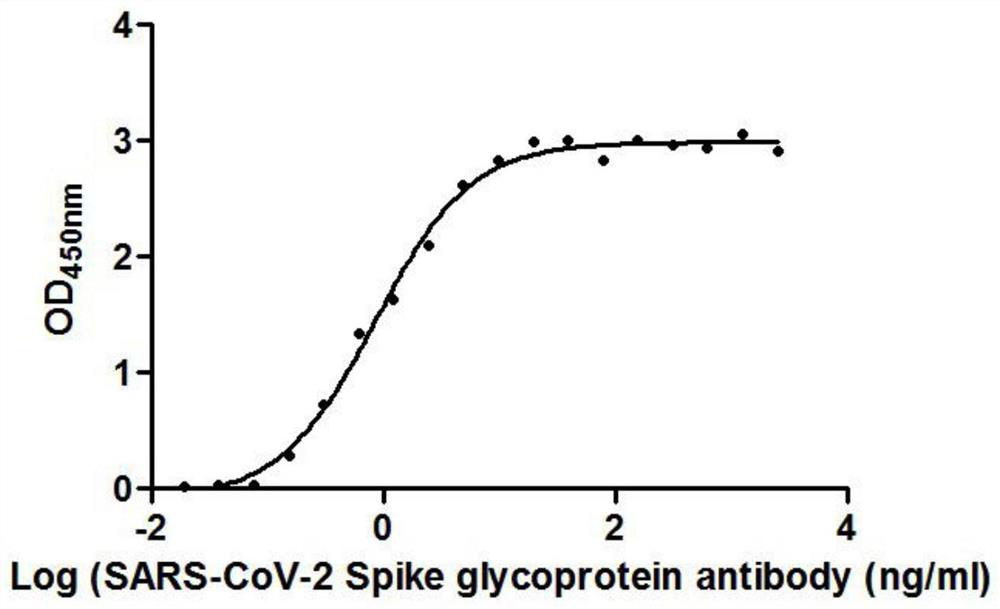
[0121] The 19 different nanobodies screened in Example 1 were combined with RBD respectively, and the EC50 value of antibody binding to RBD was detected. The RBD protein was coated on a microwell plate at 2 μg / ml. The EC50 values ​​of the 19 nanobodies are shown in Table 3. Show.

[0122] table 3

[0123] Nanobodies EC50(ng / ml) Nanobodies EC50(ng / ml) B4 91.69 G5 14.20 B6 325.24 A1 51.79 B9 153.27 F1 0.42 E3 254.45 G3 135.22 E6 5.54 C7 150.26 E7 3.26 D4 254.45 F4 50.14 H3 25.90 F5 25.33 H4 36.21 F8 76.25 H6 50.14 G2 52.19

[0124] It can be seen from the data in Table 1 that the EC50 of the 19 strains of antibodies binding to RBD ranges from 0.42ng / ml to 325.24ng / ml.

[0125] figure 1 is the binding curve of F1 antibody and RBD protein, by figure 1 It can be seen that the RBD protein is coated on the microwell plate at 2 μg / ml, an...

Embodiment 3

[0126] Example 3 Inhibition of Nanobody Binding to ACE2 Protein and RBD Protein

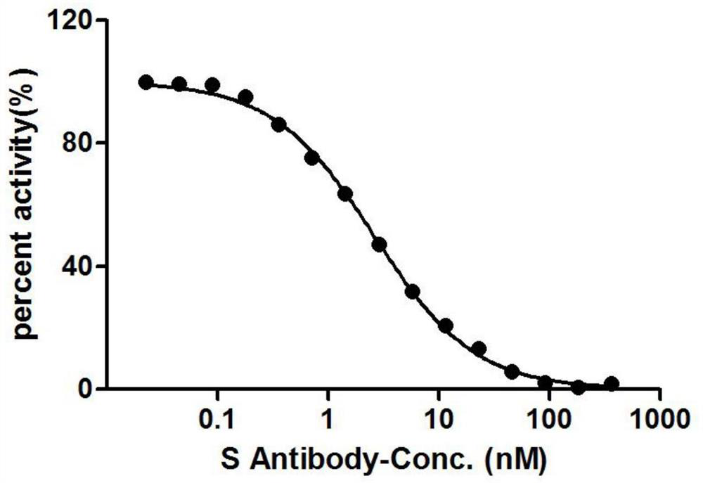
[0127] The RBD protein was coated on a microwell plate at 2 μg / ml, and the 19 different strains of nanobodies screened in Example 1 were mixed with 1 ug / ml of HRP-coupled ACE2 protein, and the OD450nm value was measured. The anti-IC50 values ​​adopted by the 19 strains of Nanobodies are shown in Table 4.

[0128] Table 4

[0129]

[0130]

[0131] Note: - indicates that the antibody has not been tested

[0132] It can be known from the data in Table 2 that all the 19 antibody strains compete with the ACE2 protein, and the IC50 ranges from 2.433nM to 100.02nM.

[0133] figure 2 It is the inhibition curve of the binding of F1 nanobody to ACE2 protein and RBD protein; the OD450nm value decreases with the increase of the concentration of F1 nanobody, indicating that there is a competitive relationship between the nanobody and ACE2 protein.


PUM


Abstract

The invention provides a nanobody against a SARS-COV-2 virus S protein RBD structure domain. The nanobody can recognize the RBD structure domain of SARS-COV-2 virus S protein, and includes one or moreof nanobody1-nanobody19. The nanobody can specifically bind to an S protein RBD region of SARS-COV-2 and block the binding of the virus to a cell receptor ACE2, and EC50 of the binding of the antibody to RBD is 0.4212ng/ml-325.24ng/ml. The nanobody competes with viral receptor ACE2 protein, and the IC50 is 2.433nm-100.02nm. The invention further provides use of the nanobody and recombinant antibody thereof in preparing drugs for inhibiting SARS-COV-2 virus infection, and preparing a SARS-COV-2 virus detection reagent or kit.

Description

technical field [0001] The invention belongs to the field of biotechnology, and relates to a nanobody against the RBD domain of the S protein of SARS-COV-2 virus and its application. Background technique [0002] The SARS-CoV-2 virus is a new type of highly pathogenic coronavirus that has ravaged the world since December 2019. The virus has 79.5% sequence similarity with the SARS virus that emerged in 2002. At the genome level, SARS-CoV-2 is 96% identical to bat coronaviruses. SARS-CoV-2 has a long incubation period of up to 14 days, so the initial stage of infection is not obvious, which makes it easier for the virus to spread, causing spread and outbreaks. In order to suppress the further spread of the epidemic, the development of rapid diagnostic kits, the development of vaccines and the preparation of neutralizing antibodies are particularly important. [0003] Coronaviruses are composed of a double-layered lipid envelope, including the spike protein, envelope protein...

Claims


Application Information

Patent Timeline
IPC IPC(8): C07K16/10C12N15/13A61K39/42A61P31/14G01N33/569
CPCA61P31/14C07K16/10C07K2317/24C07K2317/52C07K2317/565C07K2317/569C07K2317/76C07K2317/92G01N33/56983G01N2333/165G01N2469/10
Inventor 罗绍祥高霞杨旭
Owner CUSABIO TECH LLC
Why Eureka
Try Eureka