
Construction method of three-dimensional tumor model acellular derived matrix stent and application thereof

A technology of acellular matrix and construction method, which is applied in the field of construction of three-dimensional tumor model acellular porous scaffold, which can solve the problems of lack of flexibility in operation

Pending Publication Date: 2020-10-30
DALIAN UNIV OF TECH
View PDF2 Cites 2 Cited by
  • Summary
  • Abstract
  • Description
  • Claims
  • Application Information

AI Technical Summary

Problems solved by technology

Whole-organ kidney decellularized scaffolds are just to solve the problem of organ transplantation. Decellularization is aimed at the whole kidney tissue, and a sterile perfusion system must be established. The subsequent inoculation of cells to regenerate the kidney also needs to be carried out in a sterile environment, and the operation is inflexible. sex

Method used


Image

  • Construction method of three-dimensional tumor model acellular derived matrix stent and application thereof
  • Construction method of three-dimensional tumor model acellular derived matrix stent and application thereof
  • Construction method of three-dimensional tumor model acellular derived matrix stent and application thereof

Examples


Embodiment example 1

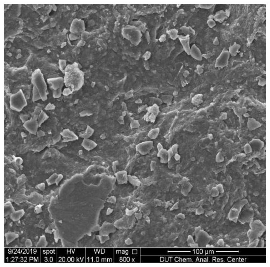
[0051] Implementation Case 1 Cross-linking and performance testing of porcine kidney-derived decellularized three-dimensional tumor model scaffold

[0052] (1) Take fresh pig kidneys from the market, wash them with water until the water has no obvious blood color, and put the washed pig kidneys in a refrigerator at -25°C for 12 hours.

[0053] (2) After it is frozen and formed, cut the pig kidney into two pieces, and remove the pig’s renal pelvis and renal fibrous capsule, and then cut the remaining kidney into a volume of 13cm 3 of small pieces.

[0054] (3) At room temperature, soak and wash the cut kidney pieces in phosphate (PBS) buffer solution with pH=7.4, and change the solution every 30 minutes for 3 times to remove blood streaks and Remove fishy smell. The cleaning process is under the action of a magnetic stirrer, in order not to damage the structure of the pig kidney small pieces, low-speed stirring is used, and the stirring rate is 8rpm.

[0055] (4) At room tem...

Embodiment example 2

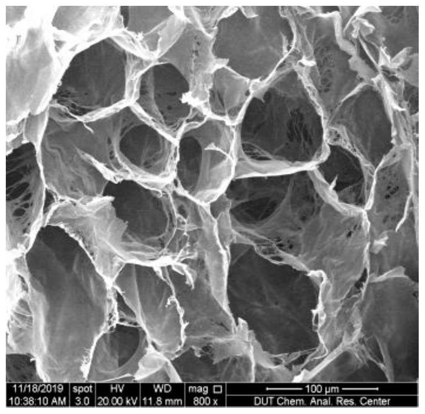
[0061] Implementation Case 2 Cross-linking and performance testing of porcine kidney-derived decellularized three-dimensional tumor model scaffold

[0062] (1) Take fresh pig kidneys from the market, wash them with water until the water has no obvious blood color, and put the washed pig kidneys in a refrigerator at -30°C for 14 hours.

[0063] (2) After it is frozen and formed, cut the pig kidney into two pieces, and remove the pig’s renal pelvis and renal fibrous capsule, and then cut the remaining kidney into a volume of 16cm 3 of small pieces.

[0064] (3) At room temperature, soak and wash the cut kidney pieces in phosphate (PBS) buffer solution with pH = 7.4, and change the solution every 30 minutes for 4 times to remove blood streaks and Remove fishy smell. In the cleaning process, under the action of a magnetic stirrer, in order not to damage the structure of the small pieces of pig kidney, low-speed stirring was used, and the stirring rate was 15 rpm.

[0065] (4) A...

Embodiment 3

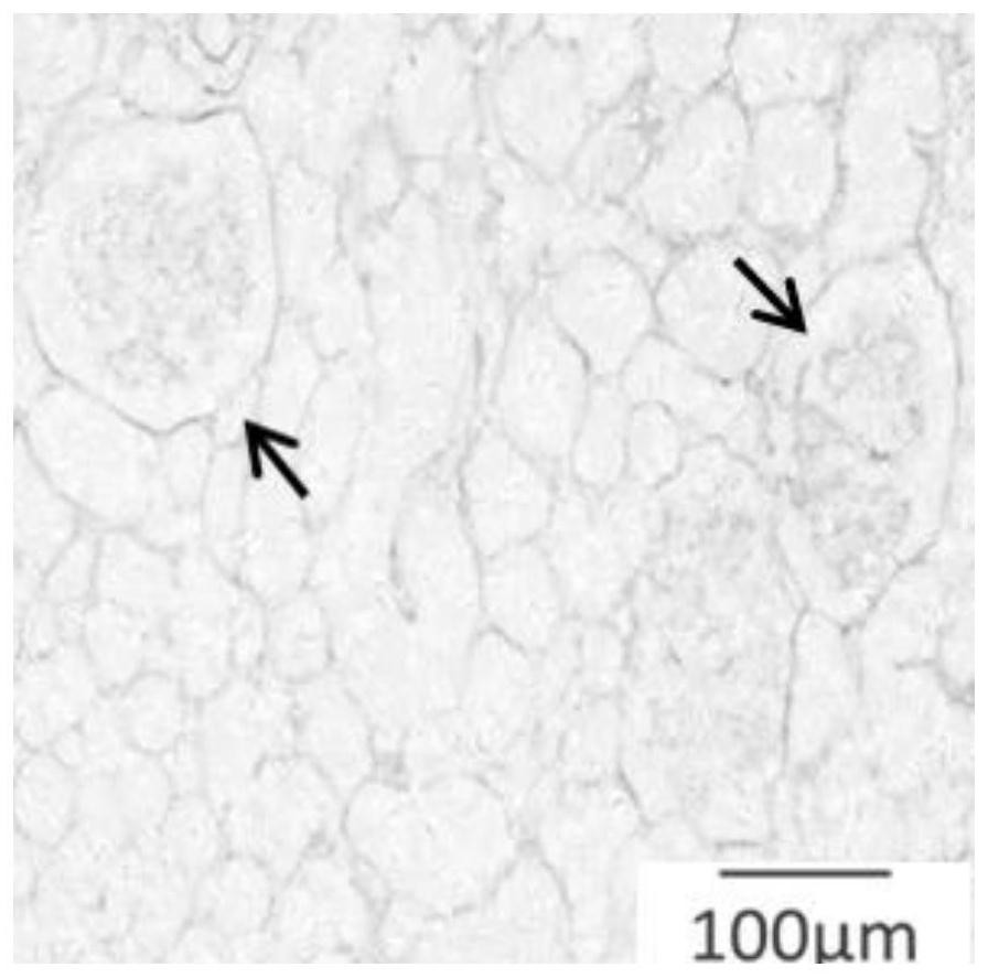
[0070] Example 3 Porcine Kidney Derived Acellular Matrix Scaffold as a Tumor Model

[0071] Take the cross-linked porcine kidney-derived acellular-derived matrix scaffold, cut off the dense outer layer on its surface with a blade, and cut into a regular scaffold with a length × width × height of 5mm × 5mm × 2mm, respectively, in order to control the variable of the scaffold volume. Better comparison of cell growth and tumor model construction under different variables of our experiments. Put the treated cross-linked scaffold into a 6-well plate filled with 75% alcohol and soak for 3 times, then replace the alcohol and put it into a sterilization table for ultraviolet sterilization overnight. After soaking in PBS for 3 hours, add DMEM medium to infiltrate, and air-dry in an ultra-clean bench. After all sterilization steps were completed, the medium was sucked away, and MCF-7 cells were inoculated for 21 days.

[0072] MCF-7 cells were inoculated on porcine kidney-derived dece...


PUM


Abstract

The invention provides a construction method of a three-dimensional tumor model acellular matrix stent and an application thereof, and belongs to the field of cytobiology and tumor tissue engineeringmaterials. The preparation method comprises the following steps: dicing pig-derived kidney tissues serving as a main material, removing cells under the action of a decellularization reagent, and preparing the pig kidney decellularization matrix porous scaffold by adopting a freeze drying technology and a chemical cross-linking technology. According to the stent prepared by the method, the matrix,blood vessels and glomerular structures of the kidney are well reserved, the biological activity of the matrix is well preserved, the utilization rate of materials is high, and the obtaining amount ofthe stent is considerable; after cancer cells are inoculated, the cells grow well on the scaffold and are distributed stereoscopically, and the cells permeate to different degrees in each layer.

Description

technical field [0001] The invention belongs to the field of cell biology and tumor tissue engineering materials, and provides a construction method and application of a three-dimensional tumor model decellularized porous scaffold. Background technique [0002] As one of the largest public health problems in the world, malignant tumor has become a common and frequently-occurring disease that seriously endangers human health. With the continuous discovery of tumor targets and anticancer drugs, choosing a model that truly simulates tumor tissue in vivo is an important means of screening effective anticancer drugs. [0003] Solid tumors grow in a three-dimensional pattern in vivo, and their occurrence and development are the result of continuous interactions between tumor cells, their microenvironment and the body. The tumor microenvironment is an important factor affecting biological behaviors such as cell secretion, adhesion, proliferation and differentiation, invasion and m...

Claims


Application Information

Patent Timeline
IPC IPC(8): C12N5/071C12N5/09C12Q1/02
CPCC12N5/0686C12N5/0693G01N33/5011C12N2533/90C12N2513/00C12N2503/02G01N2500/10
Inventor 宋克东郑乐李丽颖胡雪岩徐杰李香琴刘天庆
Owner DALIAN UNIV OF TECH
Learn More